Alternate header for print version
Advanced search
Contributors
Help
Submit
Search
menu
Cell Process
Cell Component
Cell Type
Organism
Microbial
Alzheimer's
Data Sets
Center for Research in Biological Systems
University of California, San Diego
9500 Gilman Drive
La Jolla, CA 92093-0608, USA
Voice
: (858) 534-0276
Fax
: (858) 534-7497
Email
: dorloff@ncmir.ucsd.edu
Grouped images - the images shown below are related
CIL:10094
NCBI Organism Classification
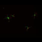
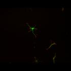
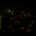
Rattus
Biological Process
developmental process
Cellular Component
cytoskeleton
Image name
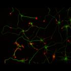
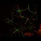
This multi-layer image shows the spatial relationship between filamentous actin ...
CIL:10095
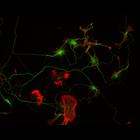
NCBI Organism Classification
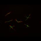
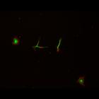
Rattus
Biological Process
developmental process
Cellular Component
cytoskeleton
Image name
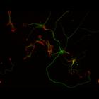
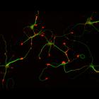
This multi-layer image shows the spatial relationship between filamentous actin ...
CIL:10096
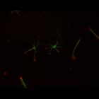
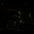
NCBI Organism Classification
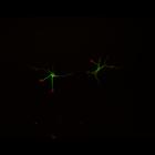
Rattus
Biological Process
developmental process
Cellular Component
cytoskeleton
Image name
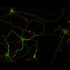
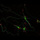
This multi-layer image shows the spatial relationship between filamentous actin ...
CIL:10097
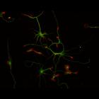
NCBI Organism Classification
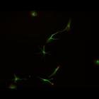
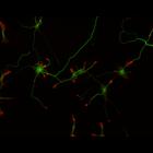
Rattus
Biological Process
developmental process
Cellular Component
cytoskeleton
Image name
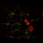
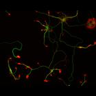
This multi-layer image shows the spatial relationship between filamentous actin ...
CIL:10098
NCBI Organism Classification
Rattus
Biological Process
developmental process
Cellular Component
cytoskeleton
Image name
This multi-layer image shows the spatial relationship between filamentous actin ...
CIL:10099
NCBI Organism Classification
Rattus
Biological Process
developmental process
Cellular Component
cytoskeleton
Image name
This multi-layer image shows the spatial relationship between filamentous actin ...
CIL:10100
NCBI Organism Classification
Rattus
Biological Process
developmental process
Cellular Component
cytoskeleton
Image name
This multi-layer image shows the spatial relationship between filamentous actin ...
CIL:10106
NCBI Organism Classification
Rattus
Biological Process
developmental process
Cellular Component
cytoskeleton
Image name
This multi-layer image shows the spatial relationship between filamentous actin ...
CIL:10107
NCBI Organism Classification
Rattus
Biological Process
developmental process
Cellular Component
cytoskeleton
Image name
This multi-layer image shows the spatial relationship between filamentous actin ...
CIL:10108
NCBI Organism Classification
Rattus
Biological Process
developmental process
Cellular Component
cytoskeleton
Image name
This multi-layer image shows the spatial relationship between filamentous actin ...
CIL:10109
NCBI Organism Classification
Rattus
Biological Process
developmental process
Cellular Component
cytoskeleton
Image name
This multi-layer image shows the spatial relationship between filamentous actin ...
CIL:10110
NCBI Organism Classification
Rattus
Biological Process
developmental process
Cellular Component
cytoskeleton
Image name
This multi-layer image shows the spatial relationship between filamentous actin ...
CIL:10111
NCBI Organism Classification
Rattus
Biological Process
developmental process
Cellular Component
cytoskeleton
Image name
This multi-layer image shows the spatial relationship between filamentous actin ...
CIL:10112
NCBI Organism Classification
Rattus
Biological Process
developmental process
Cellular Component
cytoskeleton
Image name
This multi-layer image shows the spatial relationship between filamentous actin ...
CIL:10113
NCBI Organism Classification
Rattus
Biological Process
developmental process
Cellular Component
cytoskeleton
Image name
This multi-layer image shows the spatial relationship between filamentous actin ...
CIL:10114
NCBI Organism Classification
Rattus
Biological Process
developmental process
Cellular Component
cytoskeleton
Image name
This multi-layer image shows the spatial relationship between filamentous actin ...
CIL:10115
NCBI Organism Classification
Rattus
Biological Process
developmental process
Cellular Component
cytoskeleton
Image name
This multi-layer image shows the spatial relationship between filamentous actin ...
CIL:10116
NCBI Organism Classification
Rattus
Biological Process
developmental process
Cellular Component
cytoskeleton
Image name
This multi-layer image shows the spatial relationship between filamentous actin ...
CIL:10117
NCBI Organism Classification
Rattus
Biological Process
developmental process
Cellular Component
cytoskeleton
Image name
This multi-layer image shows the spatial relationship between filamentous actin ...
CIL:10118
NCBI Organism Classification
Rattus
Biological Process
developmental process
Cellular Component
cytoskeleton
Image name
This multi-layer image shows the spatial relationship between filamentous actin ...
CIL:10119
NCBI Organism Classification
Rattus
Biological Process
developmental process
Cellular Component
cytoskeleton
Image name
This multi-layer image shows the spatial relationship between filamentous actin ...