Alternate header for print version


Licensing
Public Domain: This image is in the public domain and thus free of any copyright restrictions. However, as is the norm in scientific publishing and as a matter of courtesy, any user should credit the content provider for any public or private use of this image whenever possible. Learn more
tweet  
*CIL – Cell Image Library accession number. Please use this to reference an image.

CIL:10082*  Cite 
Description

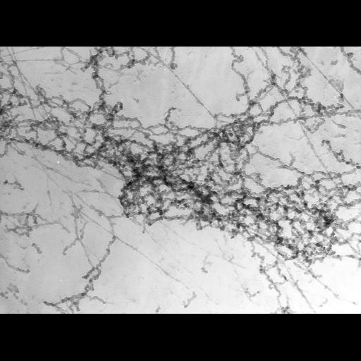
Nucleated erythrocytes from the newt Notopthahmus viridescens were spread on water, the dispersed chromatin picked up on carbon-formvar grids, fixed with paraformaldehye, critical point dried and shadowed with platinum. Images were obtained with the Wisconsin high voltage TEM at 1MEV. For this micrograph, the grid was tilted to 45 degrees. A similar micrograph tilted to 55 degrees providing a stereo pair with an oblique 3D view of the tangled and irregular chromatin fibers is grouped with this image.

Biological Sources
NCBI Organism Classification
Notopthalmus viridescence
Cell Type
erythrocyte
Cellular Component
nuclear chromatin
Attribution
Names
Hans Ris
Citation
Digital Object Identifier (DOI)
doi:10.7295/W9CIL10082
Archival Resource Key (ARK)
ark:/b7295/w9cil10082
Grouping This image is part of a group.
Imaging
Image Type
charge coupled device (CCD)
Image Mode
transmission electron microscopy (TEM)
Parameters Imaged
electron density
Source of Contrast
differences in deposition of metal shadow
Visualization Methods
shadowing and plating
Processing History
unprocessed raw data
Sample Preparation
Methods
formaldehyde fixed tissue
critical_point dried specimen
Relation To Intact Cell
spread preparation
Dimensions
Spatial Axis Image Size Pixel Size
X 3696px 1nm
Y 2742px 1nm