Alternate header for print version


Licensing
Public Domain: This image is in the public domain and thus free of any copyright restrictions. However, as is the norm in scientific publishing and as a matter of courtesy, any user should credit the content provider for any public or private use of this image whenever possible. Learn more
tweet  
*CIL – Cell Image Library accession number. Please use this to reference an image.

CIL:10703*  Cite 
Description

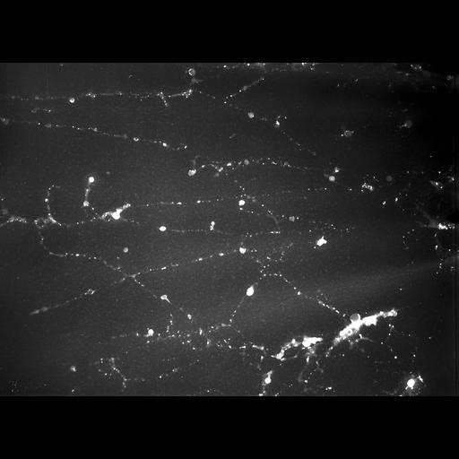
Chicken erythrocyte nuclei were allowed to disperse in low salt, centrifuged through formaldehyde onto an EM grid, stained with uranyl acetate, and examined with the Madison 1MeV TEM at 40 KX in darkfield mode. This micrograph, showing tangled chromatin fibers of varying thickness, was recorded at a tilt angle of 55 degrees. The image is grouped with one of the same sample area recorded at a 45 degree tilt. A stereo view of the pair provides an oblique 3D impression of the specimen.

Biological Sources
NCBI Organism Classification
Gallus gallus gallus
Cell Type
erythrocyte
Cellular Component
chromatin
Attribution
Names
Hans Ris
Citation
Digital Object Identifier (DOI)
doi:10.7295/W9CIL10703
Archival Resource Key (ARK)
ark:/b7295/w9cil10703
Grouping This image is part of a group.
Imaging
Image Type
transmission electron microscopy (TEM)
Image Mode
film
Parameters Imaged
electron density
Source of Contrast
differences in adsorption or binding of stain
Visualization Methods
uranyl salt
Processing History
unprocessed raw data
Sample Preparation
Methods
formaldehyde fixed tissue
Relation To Intact Cell
isolated subcellular component
Dimensions
Spatial Axis Image Size Pixel Size
X 3696px 0.5µm
Y 2686px 0.5µm