Alternate header for print version


Licensing
Attribution Non-Commercial; No Derivatives:This image is licensed under a Creative Commons Attribution, Non-Commercial, No Derivatives License. View License Deed | View Legal Code
tweet  
*CIL – Cell Image Library accession number. Please use this to reference an image.

CIL:10767*  Cite 
Description

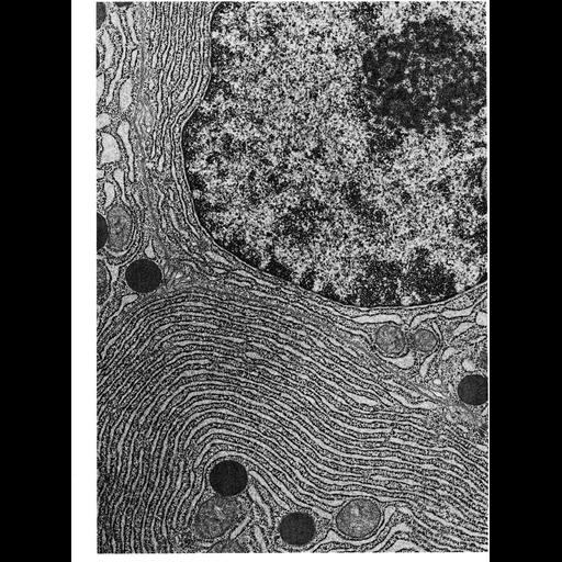
Figure 170 from Chapter 5 (Endoplasmic Reticulum) of 'The Cell, 2nd Ed.' by Don W. Fawcett M.D. Electron micrograph of rough endoplasmic reticulum (RER) in an acinar cell from the human pancreas. The RER is stacked in a cisternal structure, and studded with ribosomes. In this particular preparation, fixation conditions were modified to preserve a flocculent precipitate of newly synthesized protein in the lumen of the ER. The nucleus of the cell is in the upper right corner. Micrograph by Susmo Ito and Arthur Like. A PDF copy of the accompanying chapter is available on the ASCB’s BioEDUCATE website.

Biological Sources
NCBI Organism Classification
Homo sapiens
Cell Type
acinar cell
Cellular Component
endoplasmic reticulum
ribosome
rough endoplasmic reticulum
nucleus
endoplasmic reticulum cisternal network
Biological Context
Biological Process
translation
Attribution
Names
Susumo Ito
Arthur Like
Published
ISBN 0721635849
Link
http://www.ascb.org/bioeducate/FawcettTheCell...
Citation
Digital Object Identifier (DOI)
doi:10.7295/W9CIL10767
Archival Resource Key (ARK)
ark:/b7295/w9cil10767
Grouping This image is part of a group.
Imaging
Image Type
transmission electron microscopy (TEM)
Image Mode
illumination by electrons
detection of electrons
Parameters Imaged
electron density
Source of Contrast
stain with broad specificity
Data Qualifiers
processed data
Dimensions
Spatial Axis Image Size Pixel Size
X 950px ——
Y 1276px ——