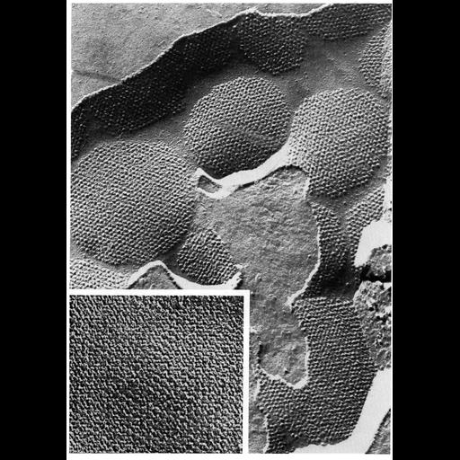
Freeze fracture image of the E-face of rat bladder lumenal membrane. The lumenal surface of these epithelial cells is composed of plaques separated by interplaque regions. Within the plaques are hexagonal arrays of subunits (about 12nm, with center-to-center spacing of 15.6nm) on the E-face of the membrane. The inset panel shows these subunits at higher magnification, prepared by deep-etching rather than fracturing after freezing. At higher magnification, each subunit within the plaque appear to be composed of a ring of six particles surrounding a channel. These plaques are distinguished from gap junctions (found on the P-face of membranes) by their location on their position instead on the E-face. These micrographs courtesy of Nicholas Severs and Marian Hicks, Figure 13 from Chapter 1 (The Cell Surface) of 'The Cell, 2nd Ed.' by Don W. Fawcett M.D. A PDF copy of the accompanying chapter is available on the ASCB's BioEDUCATE website.
With the freeze-fracture technique, tissue is rapidly frozen and cracked to shear along zones of weakness. Cleavage of membranes occurs along the hydrophobic interior of the lipid bilayer to reveal views of a "p-face" (the outwardly-facing inner half of the membrane) and an "e-face" (the inwardly-facing outer half of the membrane), and a metallic replica is made of the fractured surface. The intramembranous particles represent integral membrane proteins.
| Spatial Axis | Image Size | Pixel Size |
|---|---|---|
| X | 902px | —— |
| Y | 1272px | —— |