Alternate header for print version


Licensing
Attribution Non-Commercial; No Derivatives:This image is licensed under a Creative Commons Attribution, Non-Commercial, No Derivatives License. View License Deed | View Legal Code
tweet  
*CIL – Cell Image Library accession number. Please use this to reference an image.

CIL:10924*  Cite 
Description

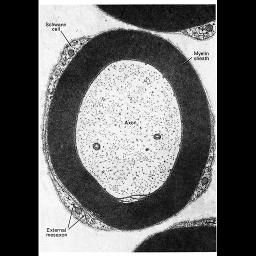
This electron micrograph shows a cross section through an axon, its myelin wrap, and the associated myelin-producing Schwann cell from the the cochlear nerve of the cat. The external mesaxon is the location where the wrapping edges of the Schwann cell meet, as the cell extends membrane that encircles the axon. This image, by Enrico Mugnaini, is Figure 14 from Chapter 1 (The Cell Surface) of 'The Cell, 2nd Ed.' by Don W. Fawcett M.D. A PDF copy of the accompanying chapter is available on the ASCB's BioEDUCATE website.

Biological Sources
NCBI Organism Classification
Felis catus
Cell Type
myelinating Schwann cell
peripheral neuron
Cellular Component
myelin sheath
axon
Attribution
Names
Enrico Mugnaini
Published
ISBN 0721635849
Link
The Cell, 2nd Ed. by D.W. Fawcett
Citation
Digital Object Identifier (DOI)
doi:10.7295/W9CIL10924
Archival Resource Key (ARK)
ark:/b7295/w9cil10924
Grouping This image is part of a group.
Imaging
Image Type
transmission electron microscopy (TEM)
Image Mode
illumination by electrons
detection of electrons
Parameters Imaged
electron density
Source of Contrast
stain with broad specificity
Visualization Methods
osmium tetroxide
Data Qualifiers
processed data
Sample Preparation
Methods
crosslinking-fixative fixed tissue
Relation To Intact Cell
microtome-sectioned tissue
Dimensions
Spatial Axis Image Size Pixel Size
X 902px ——
Y 1292px ——