Alternate header for print version


Licensing
Attribution Non-Commercial; No Derivatives:This image is licensed under a Creative Commons Attribution, Non-Commercial, No Derivatives License. View License Deed | View Legal Code
tweet  
*CIL – Cell Image Library accession number. Please use this to reference an image.

CIL:10953*  Cite 
Description

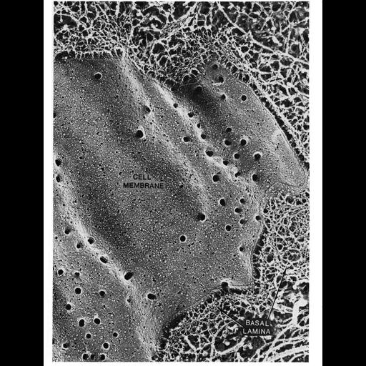
Sarcolemma of skeletal muscle prepared by a quick-freeze deep-etch method also shows the basement membrane (lamina externa, labeled here as the basal lamina) associated with the muscle fiber. This method reveals a basement membrane composed of a meshwork of fibers, some of which contact the cell membrane. The larger fibrils outside of the lamina externa (to the far lower right of the image) are composed of collagen. Image by John Heuser, Figure 29 from Chapter 1 (The Cell Surface) of 'The Cell, 2nd Ed.' by Don W. Fawcett M.D. A PDF copy of the accompanying chapter is available on the ASCB's BioEDUCATE website.

Biological Sources
Cell Type
skeletal muscle cell
Cellular Component
basement membrane
extracellular region part
plasma membrane
Attribution
Names
John Heuser
Published
ISBN 0721635849
Link
The Cell, 2nd Ed. by D.W. Fawcett
Citation
Digital Object Identifier (DOI)
doi:10.7295/W9CIL10953
Archival Resource Key (ARK)
ark:/b7295/w9cil10953
Grouping This image is part of a group.
Imaging
Image Type
transmission electron microscopy (TEM)
Image Mode
illumination by electrons
detection of electrons
Parameters Imaged
electron density
Source of Contrast
differences in deposition of metal shadow
Visualization Methods
shadowing and plating
Data Qualifiers
processed data
Sample Preparation
Methods
freeze_fracture/freeze_etch
Relation To Intact Cell
isolated subcellular component
Dimensions
Spatial Axis Image Size Pixel Size
X 966px ——
Y 1268px ——