Alternate header for print version


Licensing
Attribution Non-Commercial; No Derivatives:This image is licensed under a Creative Commons Attribution, Non-Commercial, No Derivatives License. View License Deed | View Legal Code
tweet  
*CIL – Cell Image Library accession number. Please use this to reference an image.

CIL:10976*  Cite 
Description

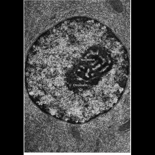
Transmission electron micrograph of nucleus showing the characteristic features of glutaraldehyde fixation introduced as a primary fixative in 1962 and since used routinely. Chromatin appears differentiated into coarse, darkly stained heterochromatin that accumulates near the nuclear periphery and the nucleolus and less darkly staining euchromatin. In contrast, chromatin appears uniformly stained with osmium tetroxide as primary fixative (for example, see CIL image 10974, in this image group). Figure 113 from Chapter 4 (Nucleus) of 'The Cell, 2nd Ed.' by Don W. Fawcett M.D. A PDF copy of the corresponding chapter is available on the ASCB's BioEDUCATE website.

Attribution
Names
Don W. Fawcett
Published
ISBN 0721635849
Published
from: Don W. Fawcett M.D ‘The Cell’ 2nd edition, W.B. Saunders Chapter 4 (Nucleus)
Link
ASCB BioEDUCATE Program
Citation
Digital Object Identifier (DOI)
doi:10.7295/W9CIL10976
Archival Resource Key (ARK)
ark:/b7295/w9cil10976
Grouping This image is part of a group.
Imaging
Image Type
film
Image Mode
transmission electron microscopy (TEM)
detection of electrons
Parameters Imaged
electron density
Source of Contrast
differences in adsorption or binding of stain
Visualization Methods
uranyl salt
osmium tetroxide
lead salt
Processing History
Image extracted from .pdf file of 'The Cell'
Data Qualifiers
processed data
Dimensions
Spatial Axis Image Size Pixel Size
X 883px ——
Y 1272px ——