Alternate header for print version


Licensing
Attribution Non-Commercial; No Derivatives:This image is licensed under a Creative Commons Attribution, Non-Commercial, No Derivatives License. View License Deed | View Legal Code
tweet  
*CIL – Cell Image Library accession number. Please use this to reference an image.

CIL:10987*  Cite 
Description

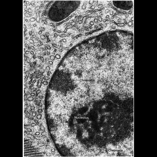
Transmission electron micrograph of glutaraldehyde fixed pancreatic acinar cell showing the characteristic features of nuclear chromatin with this preparative method. Darkly staining heterochromatin (here labeled 'chromatin') appears granular and concentrated at the nuclear periphery and nucleolus. Micrograph courtesy of Susumu Ito appears as Figure 118 from Chapter 4 (Nucleus) of 'The Cell, 2nd Ed.' by Don W. Fawcett M.D. A PDF copy of the corresponding chapter is available on the ASCB's BioEDUCATE website.

Biological Sources
NCBI Organism Classification
Unspecified
Cell Type
pancreatic acinar cell
Cellular Component
nucleus
heterochromatin
nucleolus
ribosome
rough endoplasmic reticulum
nuclear envelope
Attribution
Names
Susumu Ito
Don W Fawcett
Published
ISBN 0721635849
Published
from: Don W. Fawcett M.D ‘The Cell’ 2nd edition, W.B. Saunders Chapter 4 (Nucleus)
Link
ASCB BioEDUCATE Program
Citation
Digital Object Identifier (DOI)
doi:10.7295/W9CIL10987
Archival Resource Key (ARK)
ark:/b7295/w9cil10987
Grouping This image is part of a group.
Imaging
Image Type
film
Image Mode
transmission electron microscopy (TEM)
detection of electrons
Parameters Imaged
electron density
Source of Contrast
differences in adsorption or binding of stain
Visualization Methods
uranyl salt
osmium tetroxide
lead salt
Processing History
Image extracted from .pdf file of 'The Cell'
Data Qualifiers
processed data
Sample Preparation
Methods
tissue in plastic embedment
osmium tetroxide fixed tissue
glu
Relation To Intact Cell
microtome-sectioned tissue
Dimensions
Spatial Axis Image Size Pixel Size
X 894px ——
Y 1260px ——