Alternate header for print version


Licensing
Attribution Non-Commercial; No Derivatives:This image is licensed under a Creative Commons Attribution, Non-Commercial, No Derivatives License. View License Deed | View Legal Code
tweet  
*CIL – Cell Image Library accession number. Please use this to reference an image.

CIL:11113*  Cite 
Description

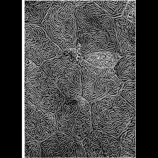
Scanning electron micrograph of the epidermal surface of the Pacific coho salmon. Ridge-like folds of the plasmalemma, called microplicae, display a labyrinth pattern across the surface, and a circumferential ridge at the borders of the polygonal-shaped cells. Image from Hawkes (1974) Cell Tissue Research, 149:147-158, reprinted with permission as Figure 42 from Chapter 2 (Specializations of the Free Surface) of 'The Cell, 2nd Ed.' by Don W. Fawcett M.D. A PDF copy of the accompanying chapter is available on the ASCB’s BioEDUCATE website.

Biological Sources
NCBI Organism Classification
Oncorhynchus kisutch
Cell Type
epidermal cell
Cellular Component
cell surface
Biological Context
Biological Process
plasma membrane organization
Attribution
Names
J. W. Hawkes
Published
ISBN 0721635849
Pubmed
PMID: 4424315
Link
Fawcett's "The Cell, 2nd Ed."
Hawkes (1974) Cell Tiss Res.
Citation
Digital Object Identifier (DOI)
doi:10.7295/W9CIL11113
Archival Resource Key (ARK)
ark:/b7295/w9cil11113
Grouping This image is part of a group.
Imaging
Image Type
scanning electron microscopy (SEM)
Image Mode
illumination by electrons
secondary_electron imaging
Parameters Imaged
secondary electron generation
Data Qualifiers
processed data
Sample Preparation
Methods
critical_point dried specimen
Dimensions
Spatial Axis Image Size Pixel Size
X 914px ——
Y 1268px ——