Alternate header for print version


Licensing
Attribution Non-Commercial; No Derivatives:This image is licensed under a Creative Commons Attribution, Non-Commercial, No Derivatives License. View License Deed | View Legal Code
tweet  
*CIL – Cell Image Library accession number. Please use this to reference an image.

CIL:11115*  Cite 
Description

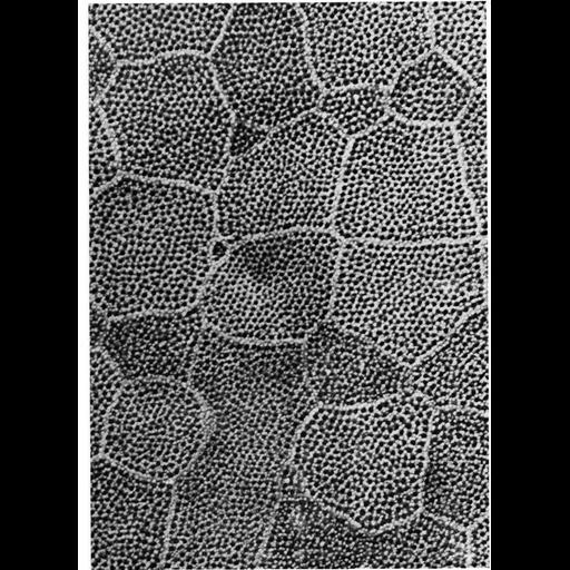
Scanning electron micrograph of the epidermal surface of lamprey larvae. A row of microvilli outline the polygonal borders between cells, while short microvilli cover the external surface in a reticular network. Image from Fahrenbach, W. (1975) J. Invest. Dermatol. 65:39-44, reprinted with permission as Figure 43 from Chapter 2 (Specializations of the Free Surface) of 'The Cell, 2nd Ed.' by Don W. Fawcett M.D. A PDF copy of the accompanying chapter is available on the ASCB’s BioEDUCATE website.

Biological Sources
NCBI Organism Classification
Entosphenus tridentatus
Cell Type
epidermal cell
Cellular Component
cell surface
external side of plasma membrane
Biological Context
Biological Process
plasma membrane organization
cell projection organization
Attribution
Names
Wolf Fahrenbach
D. D. Knutson
Published
ISBN 0721635849
Pubmed
PMID: 1151108
Link
Fawcett's "The Cell, 2nd Ed."
Fahrenbach & Knutson, J. Invest. Dermatol. 19...
Citation
Digital Object Identifier (DOI)
doi:10.7295/W9CIL11115
Archival Resource Key (ARK)
ark:/b7295/w9cil11115
Grouping This image is part of a group.
Imaging
Image Type
scanning electron microscopy (SEM)
Image Mode
illumination by electrons
secondary_electron imaging
Parameters Imaged
secondary electron generation
Data Qualifiers
processed data
Sample Preparation
Methods
critical_point dried specimen
Dimensions
Spatial Axis Image Size Pixel Size
X 927px ——
Y 1277px ——