Alternate header for print version


Licensing
Attribution Non-Commercial; No Derivatives:This image is licensed under a Creative Commons Attribution, Non-Commercial, No Derivatives License. View License Deed | View Legal Code
tweet  
*CIL – Cell Image Library accession number. Please use this to reference an image.

CIL:11123*  Cite 
Description

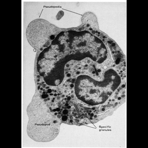
Pseudopods jut out from this polymorphonuclear leucocyte of the guinea pig to enable amoeboid motility. Organelles are initially excluded as the pseudopod first forms, but they flow in as the pseudopod enlarges. The continuous, multi-lobed nucleus appears in this field of view as two separate structures because of the thin section through the cell. Figure 45 from Chapter 2 (Specializations of the Free Surface) of 'The Cell, 2nd Ed.' by Don W. Fawcett M.D. A PDF copy of the accompanying chapter is available on the ASCB’s BioEDUCATE website.

Biological Sources
NCBI Organism Classification
Cavia porcellus
Cell Type
leukocyte
Cellular Component
pseudopodium
nucleus
Biological Context
Biological Process
cell motility
Attribution
Names
Don W. Fawcett
Published
ISBN 0721635849
Link
Fawcett's "The Cell, 2nd Ed."
Citation
Digital Object Identifier (DOI)
doi:10.7295/W9CIL11123
Archival Resource Key (ARK)
ark:/b7295/w9cil11123
Grouping This image is part of a group.
Imaging
Image Type
transmission electron microscopy (TEM)
Image Mode
illumination by electrons
detection of electrons
Parameters Imaged
electron density
Source of Contrast
stain with broad specificity
Visualization Methods
osmium tetroxide
Data Qualifiers
processed data
Dimensions
Spatial Axis Image Size Pixel Size
X 919px ——
Y 1293px ——