Alternate header for print version


Licensing
Attribution Non-Commercial; No Derivatives:This image is licensed under a Creative Commons Attribution, Non-Commercial, No Derivatives License. View License Deed | View Legal Code
tweet  
*CIL – Cell Image Library accession number. Please use this to reference an image.

CIL:11183*  Cite 
Description

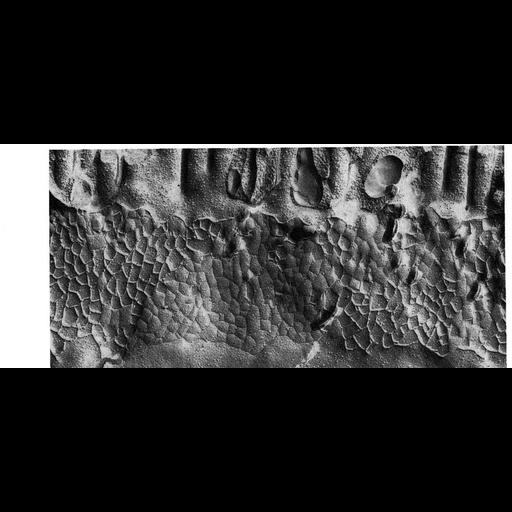
Freeze fracture replica of zonula occludens junctions from the small intestine of a Xenopus laevis tadpole. The occluding junction appears as a meshwork of ridges on the P-face or a corresponding pattern of grooves on the E-face. The geometric patterns of ridges and grooves vary depending on the epithelium. Image from Hull and Staehelin, J. Cell Biol. 68:688-704, 1976, and reprinted with permission as Figure 64 from Chapter 3 (Junctional Specializations) of 'The Cell, 2nd Ed.' by Don W. Fawcett M.D. A PDF copy of the accompanying chapter is available on the ASCB's BioEDUCATE website.

Biological Sources
NCBI Organism Classification
Xenopus laevis
Cell Type
epithelial cell
Cellular Component
tight junction
Biological Context
Biological Process
cell junction organization
Attribution
Names
Barbara Hull
Andrew Staehelin
Published
ISBN 0721635849
Pubmed
PMID:1030707
Link
The Cell, 2nd Ed. by D.W. Fawcett
Hull & Staehelin (1976) 68:688-704.
Citation
Digital Object Identifier (DOI)
doi:10.7295/W9CIL11183
Archival Resource Key (ARK)
ark:/b7295/w9cil11183
Grouping This image is part of a group.
Imaging
Image Type
transmission electron microscopy (TEM)
Image Mode
illumination by electrons
detection of electrons
Parameters Imaged
electron density
Source of Contrast
differences in deposition of metal shadow
Visualization Methods
shadowing and plating
Data Qualifiers
processed data
Sample Preparation
Methods
freeze_fracture/freeze_etch
Relation To Intact Cell
isolated subcellular component
Dimensions
Spatial Axis Image Size Pixel Size
X 993px ——
Y 436px ——