Alternate header for print version


Licensing
Attribution Non-Commercial; No Derivatives:This image is licensed under a Creative Commons Attribution, Non-Commercial, No Derivatives License. View License Deed | View Legal Code
tweet  
*CIL – Cell Image Library accession number. Please use this to reference an image.

CIL:11445*  Cite 
Description

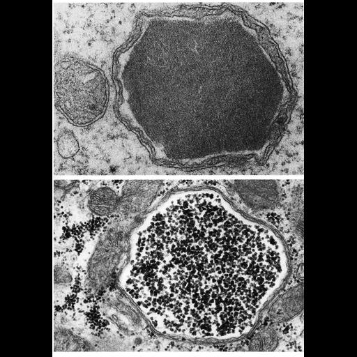
Inclusions may occur in mitochondria, either within the matrix, or as shown here, within an expanded intracristal compartment. Upper panel, a lattice of yolk crystals in an ovarian oocyte of the bullfrog Rana catesbiana; lower panel, an unusual accumulation glycogen granules in a myocardial cell from cat. Upper panel from W. Massover, J. Ultrastruct. Res., 36: 603-620, 1971). Figures 250 (upper) and 251 (lower) from Chapter 7 (Mitochondria) of 'The Cell, 2nd Ed.' by Don W. Fawcett M.D. A PDF copy of the accompanying chapter is available on the ASCB’s BioEDUCATE website.

Biological Sources
NCBI Organism Classification
Rana catesbeiana
Felis catus
Cell Type
oocyte
cardiac muscle cell
Cellular Component
mitochondrion
mitochondrial intracristal space
inclusion body
Biological Context
Biological Process
vitellogenesis
Attribution
Names
Don W. Fawcett
William Massover
Published
ISBN 0721635849
Pubmed
PMID:4329458
Link
Fawcett's "The Cell" 2nd Ed.
Citation
Digital Object Identifier (DOI)
doi:10.7295/W9CIL11445
Archival Resource Key (ARK)
ark:/b7295/w9cil11445
Grouping This image is part of a group.
Imaging
Image Type
transmission electron microscopy (TEM)
Image Mode
illumination by electrons
detection of electrons
Parameters Imaged
electron density
Source of Contrast
stain with broad specificity
Data Qualifiers
processed data
Sample Preparation
Methods
crosslinking-fixative fixed tissue
embedded tissue
Relation To Intact Cell
microtome-sectioned tissue
Dimensions
Spatial Axis Image Size Pixel Size
X 902px ——
Y 1278px ——