Alternate header for print version


Licensing
This image is copyright protected. Any public or private use of this image is subject to prevailing copyright laws. Please contact the content provider of this image for permission requests.
tweet  
*CIL – Cell Image Library accession number. Please use this to reference an image.

CIL:11503*  Cite 
Description

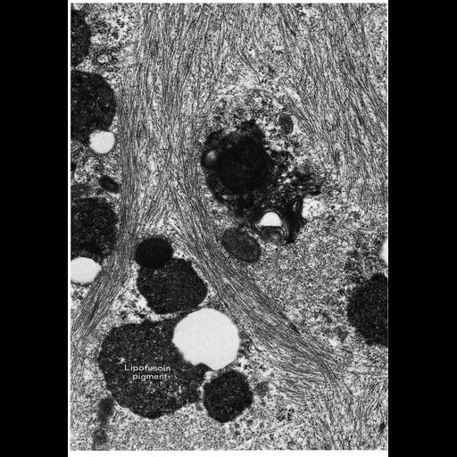
Figure 292 from Chapter 10 (Lipochrome Pigment) of 'The Cell, 2nd Ed.' by Don W. Fawcett M.D. Brown cytoplasmic inclusions identified as lipofuscin, or lipochrome pigment increase in aging cells, shown here in a Sertoli cell grown in vitro. Image by Wayne Vogl. A PDF copy of the accompanying chapter is available on the ASCB’s BioEDUCATE website.

Biological Sources
Cell Type
Sertoli cell
Cellular Component
pigment granule
Biological Context
Biological Process
aging
Attribution
Names
Don W. Fawcett
Wayne Vogl
Published
ISBN 0721635849
Link
http://www.ascb.org/bioeducate/FawcettTheCell...
Citation
Digital Object Identifier (DOI)
doi:10.7295/W9CIL11503
Archival Resource Key (ARK)
ark:/b7295/w9cil11503
Grouping This image is part of a group.
Imaging
Image Type
transmission electron microscopy (TEM)
Image Mode
illumination by electrons
detection of electrons
Parameters Imaged
electron density
Source of Contrast
stain with broad specificity
Visualization Methods
osmium tetroxide
Data Qualifiers
processed data
Dimensions
Spatial Axis Image Size Pixel Size
X 906px ——
Y 1292px ——