Alternate header for print version


Licensing
Attribution No Derivatives:This image is licensed under a Creative Commons Attribution, No Derivatives License. View License Deed | View Legal Code
tweet  
*CIL – Cell Image Library accession number. Please use this to reference an image.

CIL:11529*  Cite 
Description

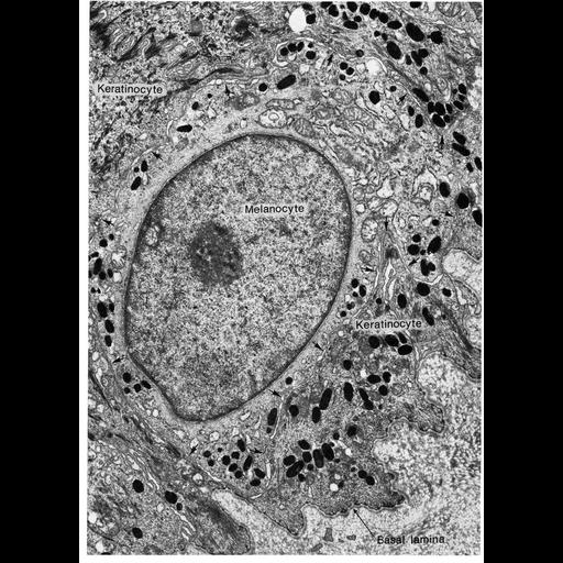
Figure 297 from Chapter 11 (Melanin Pigment) of 'The Cell, 2nd Ed.' by Don W. Fawcett M.D. Epidermal melanocyte from facial skin of an orangutan illustrates the interstitial location of a melanocyte in the basal portion of the epidermis, boundary indicated by arrows. The concentration of melanosomes is greater in the surrounding keratinocytes than in the cell body of the melanocyte. Desmosomes between epithelial cells can be observed in the upper left corner of the micrograph, but none are found at the periphery of the melanocyte. Image by George Szabo. A PDF copy of the accompanying chapter is available on the ASCB’s BioEDUCATE website.

Biological Sources
NCBI Organism Classification
Pongo pygmaeus
Cell Type
melanocyte
Cellular Component
melanosome
basal lamina
Biological Context
Biological Process
pigmentation
Attribution
Names
Don W. Fawcett
George Szabo
Published
ISBN 0721635849
Link
http://www.ascb.org/bioeducate/FawcettTheCell...
Citation
Digital Object Identifier (DOI)
doi:10.7295/W9CIL11529
Archival Resource Key (ARK)
ark:/b7295/w9cil11529
Grouping This image is part of a group.
Imaging
Image Type
transmission electron microscopy (TEM)
Image Mode
illumination by electrons
detection of electrons
Parameters Imaged
electron density
Source of Contrast
stain with broad specificity
Visualization Methods
osmium tetroxide
Data Qualifiers
processed data
Dimensions
Spatial Axis Image Size Pixel Size
X 914px ——
Y 1272px ——