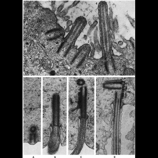
Figs. 311 & 312 from Don Fawcett's Chapter 12 (Centrioles). In ciliogenesis, newly formed single centrioles, serving as basal bodies, are arranged in rows and oriented perpendicular to the cell surface. The end that is adjacent to the membrane functions as a site of nucleation for microtubule protein which polymerizes on subunits a and b of the triplets to form the nine doublet microtubules of the axoneme. The centrioles arrive at the cell apex at different times and the polymerization of tubulin evidently begins as soon as the end of the organelle is juxtaposed to the membrane. Thus in the accompanying micrograph four developing cilia are seen in successive stages of growth. Flagella usually arise from one member of a pair of centrioles positioned at right angles. The axoneme grows out from the one perpendicular to the membrane. The lower figure on the facing page shows early stages in formation of a sperm flagellum. The differing lengths of the distal centrioles illustrated is attributable to the fact that the micrographs are not all from the same species. In one example (B) the second centriole is out of the plane of section. In sperm tail development the centrioles are involved in organizing other components in addition to the axoneme. In the most advanced stage shown (D), both centrioles are sectioned longitudinally. The thin line above the proximal centriole (*) is the anlage of the capitulum which will later articulate with the implantation fossa of the sperm nucleus. The periodic densities in the bracket (**) represent an early stage in assembly of the cross-striated columns of the connecting piece. The adjunct has begun to develop at the other end of the proximal centriole (at arrow). The centrioles thus appear to be involved in the organization of four distinct structures - axoneme, capitulum, centriolar adjunct, and connecting piece. A copy of the chapter is available on the ASCB's BioEDUCATE website.
| Spatial Axis | Image Size | Pixel Size |
|---|---|---|
| X | 922px | —— |
| Y | 1292px | —— |