Alternate header for print version


Licensing
Attribution Non-Commercial; No Derivatives:This image is licensed under a Creative Commons Attribution, Non-Commercial, No Derivatives License. View License Deed | View Legal Code
tweet  
*CIL – Cell Image Library accession number. Please use this to reference an image.

CIL:11828*  Cite 
Description

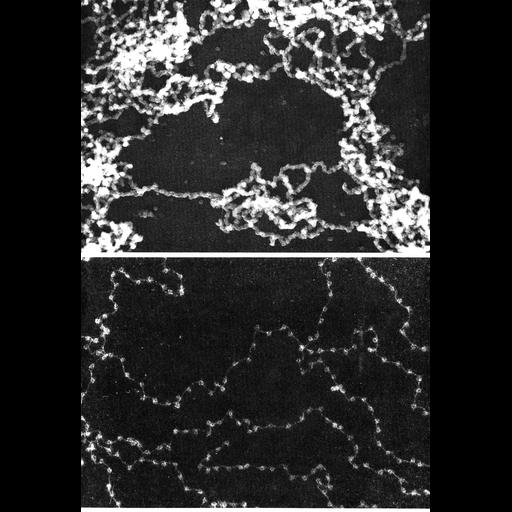
In the upper panel, transmission electron microscopy reveals a tangled mass of ~30nm diameter nuclear chromatin fibers when the contents of the nucleus from erythrocytes of the newt Notophthalmus viridescens were spread on a water surface, fixed with formaldehyde, critical point dried, and metal shadowed. The lower panel shows chromatin from the nucleated erythrocyte of the chicken dispersed in low ionic strength, high pH buffer, centrifuged through a sucrose-formaldehyde cushion onto a carbon film and stained with uranyl acetate. When fully dispersed, the 'beads-on-a-string nucleosomal organization of chromatin is clearly evident.

Technical Details

Figures 121 (upper panel, provided by Hans Ris) and 122 (lower, provided by Ada Olons) from Chapter 4 (Nucleus) of 'The Cell, 2nd Ed.' by Don W. Fawcett M.D. A PDF copy of the chapter is available on the ASCB's BioEDUCATE website.

Biological Sources
NCBI Organism Classification
Notophthalmus viridescens
Gallus gallus
Cell Type
erythrocyte
Cellular Component
chromatin
nucleosome
Biological Context
Biological Process
nucleus organization
chromatin organization
Attribution
Names
Hans Ris
Ada Olins
Don W Fawcett
Published
ISBN 0721635849
Published
from: Don W. Fawcett M.D ‘The Cell’ 2nd edition, W.B. Saunders Chapter 4 (Nucleus)
Link
ASCB BioEDUCATE Program
Citation
Digital Object Identifier (DOI)
doi:10.7295/W9CIL11828
Archival Resource Key (ARK)
ark:/b7295/w9cil11828
Grouping This image is part of a group.
Imaging
Image Type
film
Image Mode
transmission electron microscopy (TEM)
detection of electrons
Parameters Imaged
electron density
Source of Contrast
differences in adsorption or binding of stain
differences in deposition of metal shadow
Visualization Methods
uranyl salt
Processing History
Image extracted from .pdf file of 'The Cell'
Data Qualifiers
processed data
Dimensions
Spatial Axis Image Size Pixel Size
X 865px ——
Y 1268px ——