Alternate header for print version


Licensing
Public Domain: This image is in the public domain and thus free of any copyright restrictions. However, as is the norm in scientific publishing and as a matter of courtesy, any user should credit the content provider for any public or private use of this image whenever possible. Learn more
tweet  
*CIL – Cell Image Library accession number. Please use this to reference an image.

CIL:11848*  Cite 
Description

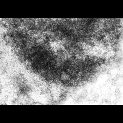
Dividing cells from the liquid endosperm of the lily Haemanthus katherinae were fixed with paraformaldehye, embedded in epoxy, and 0.5 micrometer sections prepared. Images were obtained with the Wisconsin high voltage TEM at 1MEV. For this micrograph of interphase, the grid was tilted to 60 degrees. A similar micrograph tilted to 40 degrees is grouped with this image, making a stereo pair that provides an oblique 3D view.

Biological Sources
NCBI Organism Classification
Haemanthus katharinae
Cell Type
endosperm
Cellular Component
nuclear chromosome
Biological Context
Biological Process
mitosis
Attribution
Names
Hans Ris
Citation
Digital Object Identifier (DOI)
doi:10.7295/W9CIL11848
Archival Resource Key (ARK)
ark:/b7295/w9cil11848
Grouping This image is part of a group.
Imaging
Image Type
film
Image Mode
transmission electron microscopy (TEM)
Parameters Imaged
electron density
Source of Contrast
elastic scattering of electrons
Visualization Methods
uranyl salt
Processing History
unprocessed raw data
Sample Preparation
Methods
formaldehyde fixed tissue
Relation To Intact Cell
microtome-sectioned tissue
Dimensions
Spatial Axis Image Size Pixel Size
X 3873px 2nm
Y 2673px 2nm