Alternate header for print version


Licensing
Public Domain: This image is in the public domain and thus free of any copyright restrictions. However, as is the norm in scientific publishing and as a matter of courtesy, any user should credit the content provider for any public or private use of this image whenever possible. Learn more
tweet  
*CIL – Cell Image Library accession number. Please use this to reference an image.

CIL:12035*  Cite 
Description

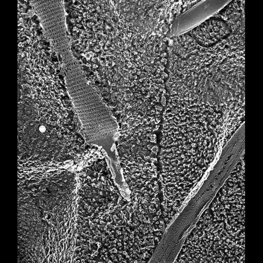
A high resolution image of portion of an isolated cilium exposing two rows of outer dynein arms arising from subfiber A of each doublet. The heavy chains of the dynein complex are linked to the microtubule by stems that can be seen in this micrograph. Thin protrusions link the dynein heavy chains to the adjacent doublet. Much of this cilium is still covered by the ciliary membrane. Also included in this image is an extruded trichocyst. TEM taken on 6/7/88 by C. Schroeder with Zeiss 10A operating at 80kV. Neg. 19,800X. The raw film was scanned with an Epson Perfection V750 Pro. This image is best used for quantitative analysis.

Biological Sources
NCBI Organism Classification
Paramecium multimicronucleatum
Cell Type
cell by organism
eukaryotic cell
Eukaryotic Protist
Ciliated Protist
Cellular Component
cilium
cilium axoneme
axonemal dynein complex
trichocyst
Biological Context
Biological Process
axonemal dynein complex assembly
trichocyst discharge
Attribution
Names
Richard Allen (University of Hawaii)
C. Schroeder
Link
http://www5.pbrc.hawaii.edu/allen/
Citation
Digital Object Identifier (DOI)
doi:10.7295/W9CIL12035
Archival Resource Key (ARK)
ark:/b7295/w9cil12035
Grouping This image is part of a group.
Dimensions
Spatial Axis Image Size Pixel Size
X 3263px 1nm
Y 3770px 1nm