Alternate header for print version


Licensing
Public Domain: This image is in the public domain and thus free of any copyright restrictions. However, as is the norm in scientific publishing and as a matter of courtesy, any user should credit the content provider for any public or private use of this image whenever possible. Learn more
tweet  
*CIL – Cell Image Library accession number. Please use this to reference an image.

CIL:12303*  Cite 
Description

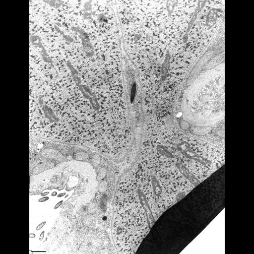
The division furrow of a dividing Didinium. A thick layer of epiplasm coated on the cytosolic side by mitochondria forms the constricting ring in the furrow. As usual mucocysts lie between the epiplasm and the pellicular membranes. The new cytopharynx in the opisthe already has lamellae and the alveolus seems to be absent from this region. The macronucleus is constricted in the furrow and elongated nucleoli are stretched out along microtubules are evident. TEM taken on 5/21/69 by R. Allen with Philips 300 operating at 60kv. Neg. 5,000X. Bar = 1µm. The negative was printed to paper and the image was scanned to Photoshop. This digitized image is available for qualitative analysis. A high resolution image is in the library (CIL:38865) and can be used for qualitative analysis. Standard glutaraldehyde fixation followed by osmium tetroxide, dehydrated in alcohol and embedded in an epoxy resin. Microtome sections prepared at approximately 75nm thickness. Additional information available at (http://www5.pbrc.hawaii.edu/allen/).

Biological Sources
NCBI Organism Classification
Didinium nasutum
Cell Type
cell by organism
eukaryotic cell
Eukaryotic Protist
Ciliated Protist
Cell Line
Carolina Biological Supply Company, NC, U.S.A.
Cellular Component
cell division site
cleavage furrow
nuclear division
extrusomes
nucleolar chromatin
Biological Context
Biological Process
cell division
cortical microtubule organization
nuclear division
nuclear microtubules
nucleolar organization
Molecular Function
structural constituent of cytoskeleton
structural molecule activity
nucleolar organization
Attribution
Names
Richard Allen
Link
http://www5.pbrc.hawaii.edu/allen/
Citation
Digital Object Identifier (DOI)
doi:10.7295/W9CIL12303
Archival Resource Key (ARK)
ark:/b7295/w9cil12303
Grouping This image is part of a group.
Imaging
Image Type
transmission electron microscopy (TEM)
illumination by electrons
Image Mode
detection of electrons
film
Parameters Imaged
electron density
Source of Contrast
stain with broad specificity
Visualization Methods
stain with broad specificity
osmium tetroxide
uranyl salt
lead salt
Processing History
recorded image
film
Print from negative scanned to Photoshop.
Data Qualifiers
processed data
suitable for spatial measurements
Dimensions
Spatial Axis Image Size Pixel Size
X 2912px ——
Y 3781px ——