Alternate header for print version


Licensing
Public Domain: This image is in the public domain and thus free of any copyright restrictions. However, as is the norm in scientific publishing and as a matter of courtesy, any user should credit the content provider for any public or private use of this image whenever possible. Learn more
tweet  
*CIL – Cell Image Library accession number. Please use this to reference an image.

CIL:12349*  Cite 
Description

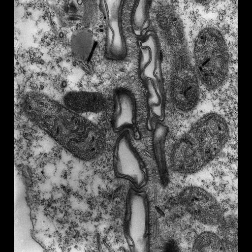
High resolution view of the cytoproct ridge at higher magnification. The unique system of thick fibers extends from each side of the ridge toward the center of the ridge. These thickened fibers are not found in any other part of the cell’s surface. Also extending from the ridge tip are single microtubules that extend down into the cytosol. These microtubules may make contact with the surface of the approaching vacuole’s membrane and help to pull the vacuole membrane and the plasma membrane together to promote their fusion. The role of the thickened fibers is unclear, they may cross link to prevent the cytoproct from an untimely opening under stress. A few vesicles get trapped in the ridge. TEM taken on 6/1/84 by R. Allen with Zeiss 10A 80kV. Neg. 19,800X. The raw film was scanned with an Epson Perfection V750 Pro. This image is best used for quantitative analysis. Standard glutaraldehyde fixation followed by osmium tetroxide, dehydrated in alcohol and embedded in an epoxy resin. Microtome sections prepared at approximately 75nm thickness. Additional information available at (http://www5.pbrc.hawaii.edu/allen/).

Biological Sources
NCBI Organism Classification
Paramecium multimicronucleatum
Cell Type
cell by organism
eukaryotic cell
Eukaryotic Protist
Ciliated Protist
Cellular Component
cytoproct
cell cortex
Biological Context
Biological Process
cytoproct organization
cortex organization
Attribution
Names
Richard Allen (University of Hawaii)
Link
http://www5.pbrc.hawaii.edu/allen/
Citation
Digital Object Identifier (DOI)
doi:10.7295/W9CIL12349
Archival Resource Key (ARK)
ark:/b7295/w9cil12349
Grouping This image is part of a group.
Imaging
Image Type
transmission electron microscopy (TEM)
illumination by electrons
Image Mode
detection of electrons
film
Parameters Imaged
electron density
Source of Contrast
stain with broad specificity
Visualization Methods
stain with broad specificity
osmium tetroxide
uranyl salt
lead salt
Processing History
recorded image
film
unprocessed raw data
Data Qualifiers
raw, unprocessed data
suitable for spatial measurements
Dimensions
Spatial Axis Image Size Pixel Size
X 3675px 1nm
Y 4128px 1nm