Alternate header for print version


Licensing
Public Domain: This image is in the public domain and thus free of any copyright restrictions. However, as is the norm in scientific publishing and as a matter of courtesy, any user should credit the content provider for any public or private use of this image whenever possible. Learn more
tweet  
*CIL – Cell Image Library accession number. Please use this to reference an image.

CIL:12628*  Cite 
Description

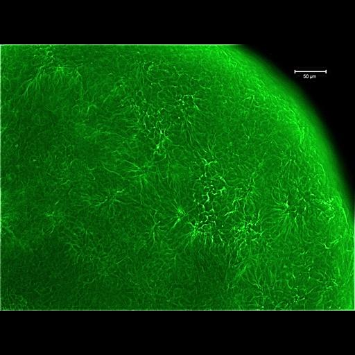
Fluorescent image of a mouse oocyte labeled with an antibody to tubulin, which is shown localized in the cortex of the oocyte. The sample was fixed in paraformaldehyde for subsequent immunolabeling with an antibody to tubulin and a secondly antibody conjugated to Alexa 488, which labels the microtubules green. The oocyte was imaged using a Nikon Eclipse inverted microscope and the original image was deconvoluted using Nikon's Elements software.

Biological Sources
NCBI Organism Classification
Mus musculus
Cell Type
oocyte
Cellular Component
cell cortex
microtubule cytoskeleton
Attribution
Names
Sabrina Lin
Prue Talbot
Citation
Digital Object Identifier (DOI)
doi:10.7295/W9CIL12628
Archival Resource Key (ARK)
ark:/b7295/w9cil12628
Imaging
Image Type
recorded image
Image Mode
fluorescence microscopy
Parameters Imaged
fluorescence emission
Source of Contrast
distribution of a specific protein
Visualization Methods
Alexa Fluor 488
primary antibody plus labeled secondary antibody
Processing History
deconvolution
Data Qualifiers
processed data
Sample Preparation
Methods
formaldehyde fixed tissue
Relation To Intact Cell
whole mounted tissue
Dimensions
Spatial Axis Image Size Pixel Size
X 1600px 0.345µm
Y 1200px 0.345µm