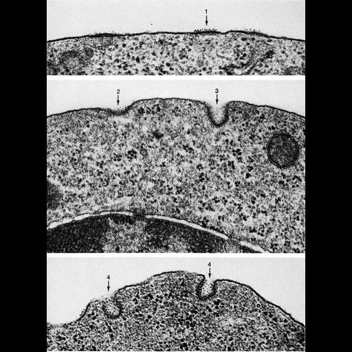
Stages of coated pit vesicle invagination during micropinocytosis. Each frame is a portion of the surface of polychromatophilic erythroblasts from guinea pig bone marrow and various stages of vesicle formation beginning with initial thickening of the membrane (1), depression (2), subsequent deepening of the pit (3), and narrowing of the neck (4). Regions of thickened membrane at sites of invagination are enhanced in this preparation by ferritin, which binds to the external region of the thickened membrane. From Fawcett (1965) J. Histochem. Cytochem. 13:75-91, reprinted with permission as Figures 54, 55, and 56 (upper, middle, lower respectively) from Chapter 2 (Specializations of the Free Surface) of 'The Cell, 2nd Ed.' by Don W. Fawcett M.D. A PDF copy of the accompanying chapter is available on the ASCB’s BioEDUCATE website.
| Spatial Axis | Image Size | Pixel Size |
|---|---|---|
| X | 1156px | —— |
| Y | 1574px | —— |