Alternate header for print version


Licensing
Public Domain: This image is in the public domain and thus free of any copyright restrictions. However, as is the norm in scientific publishing and as a matter of courtesy, any user should credit the content provider for any public or private use of this image whenever possible. Learn more
tweet  
*CIL – Cell Image Library accession number. Please use this to reference an image.

CIL:13154*  Cite 
Description

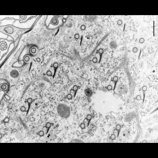
A tangential section through the cell’s surface show the pairs of basal bodies in cross section. The ends of kinetodesmal fibers point anteriorly in the cell, the postciliary ribbons arise from the posterior right side of the posterior basal body of a pair and transverse ribbons arise from the left side of both basal bodies of a pair. The more anterior basal body of a pair is linked to the posterior basal body by a striated fiber and to the adjacent kinetodesmal fiber by a short filamentous connection (arrowhead). Parasomal sacs, infraciliary lattice, striated band, and trichocyst tips are evident. TEM taken on 1/12/73 by R. Allen with Hitachi HU11A operating at 75kV. Neg. 14,900X. The negative was printed to paper and the image was scanned to Photoshop. This digitized image is available for qualitative analysis. An unprocessed, high resolution version of this image (CIL:12031) is in the library and available for quantitative analysis. Standard glutaraldehyde fixation followed by osmium tetroxide, dehydrated in alcohol and embedded in an epoxy resin. Microtome sections prepared at approximately 75nm thickness. Additional information available at (http://www5.pbrc.hawaii.edu/allen/).

Biological Sources
NCBI Organism Classification
Paramecium multimicronucleatum
Cell Type
cell by organism
eukaryotic cell
Eukaryotic Protist
Ciliated Protist
Cellular Component
cell cortex
microtubule basal body
kinetodesmal fiber
microtubule
trichocyst
Attribution
Names
Richard Allen (University of Hawaii)
Link
http://www5.pbrc.hawaii.edu/allen/
Citation
Digital Object Identifier (DOI)
doi:10.7295/W9CIL13154
Archival Resource Key (ARK)
ark:/b7295/w9cil13154
Grouping This image is part of a group.
Imaging
Image Type
transmission electron microscopy (TEM)
illumination by electrons
Image Mode
detection of electrons
film
Parameters Imaged
electron density
Source of Contrast
stain with broad specificity
Visualization Methods
stain with broad specificity
osmium tetroxide
uranyl salt
lead salt
Processing History
recorded image
film
Print from negative scanned for Photoshop.
Data Qualifiers
processed data
Dimensions
Spatial Axis Image Size Pixel Size
X 2595px ——
Y 2103px ——