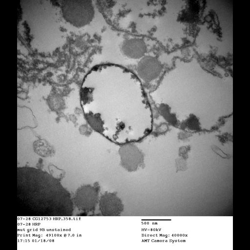
Transmission electron micrograph of Drosophila nephrocyte garland cell from ema[1] mutant pulsed with the fluid-phase endocytic tracer HRP, detected by chromogenic staining (DAB). HRP reaction product in most labeled endosome from ema[1] mutant cells does not fill the luminal space as in wild type, but instead is concentrated along the limiting membrane and in intraluminal clusters, which often extend from the limiting membrane. Third instar larvae were dissected in ice-cold PBS, equilibrated in HL-3 buffer at room temperature, incubated in 0.7% HRP (Sigma-Aldrich grade VI) for 5 min at room temperature, rinsed thoroughly, and chased for 10 min at room temperature. Samples were fixed for 1 h at 4°C in 2% paraformaldehyde, 2% glutaraldehyde, and 1% tannic acid in 0.1 M cacodylic acid buffer, pH 7.2, and processed for DAB reaction as described previously (Kosaka and Ikeda, 1983). Garland cells were dissected out and fixed for 1 h at 4°C in 2% paraformaldehyde, 2% glutaraldehyde, and 1% tannic acid in 0.1 M cacodylic acid buffer, pH 7.2. Then, the samples were postfixed in 1% OsO4 in 0.1 M cacodylic acid buffer, pH 7.2, for 1 h at room temperature, stained en bloc with 1% uranyl acetate, dehydrated in a grade series of ethanol and propylene oxide, and embedded in epon resin (Electron Microscopy Sciences). Blocks were sectioned in an ultramicrotome (RMC Products) at ∼70-nm thickness with a Delaware Diamond knife and post-stained for 1 h in Reynolds lead citrate and uranyl acetate. Electron micrographs were taken on a transmission electron microscope (H-7500; Hitachi) using an AMT camera system. Mag: 40000x. Image corresponds to Figure 4C, bottom panel in Kim et al. J Cell Biol. 188: 717-734. 2010.
| Spatial Axis | Image Size | Pixel Size |
|---|---|---|
| X | 1024px | —— |
| Y | 1184px | —— |