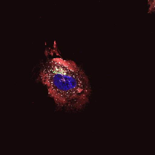
Helix Pomatia Lectin (HPL) (gray) staining is present in diffuse structures that stain with an ER marker, PDI (protein disulfide isomerase) (red) in HeLa cells upon EGF treatment for 4h. The image includes the Golgi (Giantin) (green) HPL binds various glycans but the Tn antigen in particular. The Tn antigen refers to terminal α-linked N-acetyl galactosamine residues (GalNAc) linked to Ser or Thr residues. HeLa cells were serum starved overnight in DME (noFBS) and then treated with human recombinant EGF (100 ng/ml; Sigma-Aldrich) for 4h. Cells were fixed for 10 min (4% paraformaldehyde) and permeabilized (0.2% Triton X-100). Primary antibody staining followed the manufacturer’s instructions. Cells were subsequently stained for 15–30 min with secondary Alexa Fluor–conjugated antibodies (Alexa 488 for anti-Giantin, Alexa 594 for anti PDI). Hoechst (blue) and Alexa 647-conjugated-HPL were added during secondary antibody incubations. Cells were mounted onto glass slides using FluorSave (Merck) and imaged at room temperature using an inverted FluoView confocal microscope (model IX81; Olympus) with fluorescence excitation at 488 nm, 561 nm and 633 nm and either a 60x objective (U Plan Super Apochromatic [UPLSAPO]; NA 1.35) or 100x objective (UPLSAPO; NA 1.40) using Immersol oil. Microscope coupled with a CCD camera (model FVII). Images were acquired and processed using Olympus FV10-ASW software. Image corresponds to Fig 4D in J Cell Biol. 189: 843-858. 2010. Images in Fig 4 include CIL#s 13555, 13556, 13557, 13558.
| Spatial Axis | Image Size | Pixel Size |
|---|---|---|
| X | 1024px | 0.124µm |
| Y | 1024px | 0.124µm |
| Z | 1px | 1.09µm |