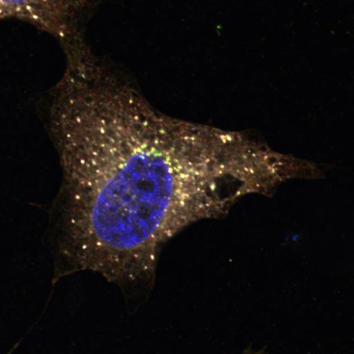
GalNac-T1 (green) is in dispersed punctate structures that stain positive for COP-I beta1 (red) after EGF treatment of HeLa cells for 4h. These data suggest that GalNAc-Ts are being redistributed from the Golgi apparatus to the ER by a process that requires COP-I vesicles. Helix Pomatia Lectin (HPL) binds various glycans but the Tn antigen in particular. The Tn antigen refers to terminal α-linked N-acetyl galactosamine residues (GalNAc) linked to Ser or Thr residues. HeLa cells were serum starved overnight in DME (noFBS) and then treated with human recombinant EGF (100 ng/ml; Sigma-Aldrich) for 4h. Cells were fixed for 10 min (4% paraformaldehyde) and permeabilized (0.2% Triton X-100). Primary antibody staining followed the manufacturer’s instructions. Cells were subsequently stained for 15–30 min with secondary Alexa Fluor–conjugated antibodies (Alexa 488 for anti-GalNac-T1, Alexa 594 for anti-COP-I beta1). Hoechst (blue) and Alexa 647-conjugated-HPL were added during secondary antibody incubations. Cells were mounted onto glass slides using FluorSave (Merck) and imaged at room temperature using an inverted FluoView confocal microscope (model IX81; Olympus) with fluorescence excitation at 488 nm, 561 nm, and 633 nm and either a 60x objective (U Plan Super Apochromatic [UPLSAPO]; NA 1.35) or 100x objective (UPLSAPO; NA 1.40) using Immersol oil. Microscope coupled with a CCD camera (model FVII). Images were acquired and processed using Olympus FV10-ASW software. Image corresponds to Fig 6D, in J Cell Biol. 189: 843-858. 2010. Images in Fig 6 include CIL#s 13563, 13564, 13565, 13566, 13567, 13568, 13569, 13570.
| Spatial Axis | Image Size | Pixel Size |
|---|---|---|
| X | 1024px | 0.062µm |
| Y | 1024px | 0.062µm |
| Z | 1px | 11.03µm |