Alternate header for print version


Licensing
Attribution Non-Commercial Share Alike:This image is licensed under a Creative Commons Attribution, Non-Commercial Share Alike License. View License Deed | View Legal Code
tweet  
*CIL – Cell Image Library accession number. Please use this to reference an image.

CIL:13592*  Cite 
Description

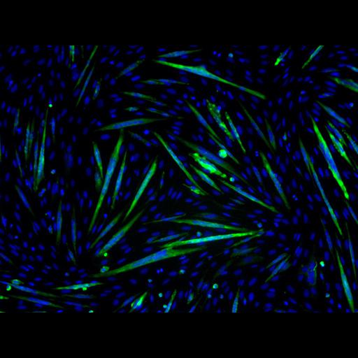
The C2C12 cell line, a mouse myoblast line, was used here to study the regulatory factors in myogenic differentiation. After cultured in differentiation medium for 3 days, these cells differentiated into myotubes (Green) containing multiple nuclei (Blue). The C2C12 cells stably expressing FLAG-tagged rapamycin-resistant, kinase-inactive (RR/KI) mTOR were induced to differentiation in the presence of 50nM rapamycin for 3 days. The C2C12 cells differentiated, but arrested at the nascent myotube stage. This image is the control of different treatments in Figure 9B from JCB 189: 1157-1169, 2010. See also CIL: 13593, 13594, 13601.

Technical Details

The image was taken by leica DMI 4000B microscope equipped with a QImaging RETIGA Exi camera and Leica 10X/0.22 lens. Myosin heavy chain (MHC) expressed in myotubes was labeled by MF-20 mouse antibody followed by FITC-anti-mouse IgG secondary antibody. Nuclei were stained by DAPI. MHC and nuclei were pseudocolored in green and blue, respectively.

Biological Sources
NCBI Organism Classification
Mus musculus
Cell Type
myoblast
Cell Line
C2C12
Cellular Component
nucleus
muscle myosin complex
Biological Context
Biological Process
myoblast fusion
Molecular Function
mTOR inhibition
Attribution
Names
Yuting Sun
Yejing Ge
Jenny Drnevich
Yong Zhao
Mark Band
Jie Chen
Published
JCB 189: 1157-1169, 2010
Pubmed
20566686
Citation
Digital Object Identifier (DOI)
doi:10.7295/W9CIL13592
Archival Resource Key (ARK)
ark:/b7295/w9cil13592
Grouping This image is part of a group.
Imaging
Image Type
recorded image
Image Mode
fluorescence microscopy
Parameters Imaged
fluorescence emission
Source of Contrast
compartmentalization of stain or label
distribution of epitope
Visualization Methods
Fluorescein (FITC)
4',6-diamidino-2-phenylindole (DAPI)
primary antibody plus labeled secondary antibody
Processing History
brightness and contrast adjusted
Data Qualifiers
processed data
suitable for spatial measurements
Sample Preparation
Methods
formaldehyde fixed tissue
permeabilized tissue
Relation To Intact Cell
dispersed cells in vitro
Dimensions
Spatial Axis Image Size Pixel Size
X 1392px 0.645µm
Y 1040px 0.645µm