Alternate header for print version


Licensing
Attribution Non-Commercial Share Alike:This image is licensed under a Creative Commons Attribution, Non-Commercial Share Alike License. View License Deed | View Legal Code
tweet  
*CIL – Cell Image Library accession number. Please use this to reference an image.

CIL:13681*  Cite 
Description

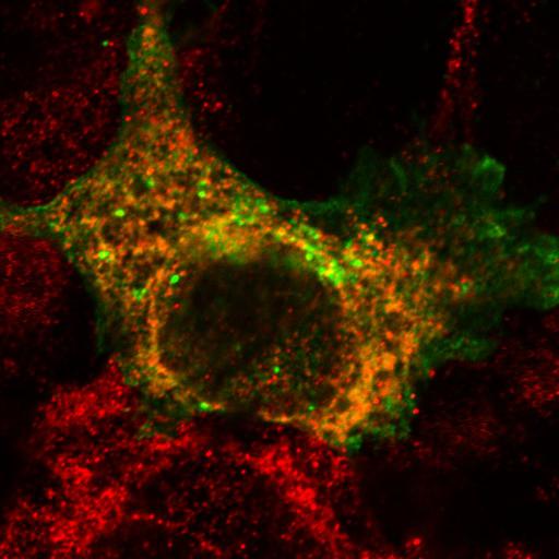
To study the cellular consequences of misfolding of plasma membrane proteins, a transmembrane model protein that was constitutively targeted to the plasma membrane was developed for use as a reporter molecule. The reporter consisted of a C-terminally truncated CD4, incorporating a flexible cytoplasmic linker (CD4tl), fused to the N-terminal DNA-binding domain of the wild type (wt) bacteriophage lambda repressor (CD4tl-lambda) or L57C mutant repressor (CD4tl-lambdaC). The CD4tl-lambdaC cytosolic domain was largely in native state at 26°C but predominantly nonnative state at 37°C thus allowing the use of thermal shifts to follow the fate of unfolded proteins. In this image, one of six of a group from Figure 1 of Pirjo et al., JCB 2010, the expression of the mutant plasma membrane reporter, CD4tl-lambdaC, at 37 degrees is seen with CD4 antibody (green) in a detergent-permeabilized COS7 cell. The intracellular processing of this nonnative, thermally unfolded protein nearly completely limits its targeting to the plasma membrane. This cell is co-labeled with an ER marker, calreticulin antibody (red).

Biological Sources
NCBI Organism Classification
Chlorocebus aethiops
Cell Line
COS-7
Cellular Component
integral to plasma membrane
endoplasmic reticulum
Biological Context
Biological Process
protein targeting to plasma membrane
Attribution
Names
Pirjo M. Apaja
Haijin Xu
Gergely L. Lukacs
Published
JCB 2010, 191:553-570
Pubmed
20974815
Citation
Digital Object Identifier (DOI)
doi:10.7295/W9CIL13681
Archival Resource Key (ARK)
ark:/b7295/w9cil13681
Grouping This image is part of a group.
Imaging
Image Type
recorded image
Image Mode
confocal microscopy
Parameters Imaged
fluorescence emission
Source of Contrast
distribution of a specific protein
Visualization Methods
Alexa Fluor 488
Alexa Fluor 555
Processing History
unprocessed raw data
Data Qualifiers
raw, unprocessed data
Sample Preparation
Methods
crosslinking-fixative fixed tissue
detergent permeabilized
Relation To Intact Cell
dispersed cells in vitro
Dimensions
Spatial Axis Image Size Pixel Size
X 1024px 47.8nm
Y 1024px 47.8nm