Alternate header for print version


Licensing
Attribution Non-Commercial Share Alike:This image is licensed under a Creative Commons Attribution, Non-Commercial Share Alike License. View License Deed | View Legal Code
tweet  
*CIL – Cell Image Library accession number. Please use this to reference an image.

CIL:13714*  Cite 
Description

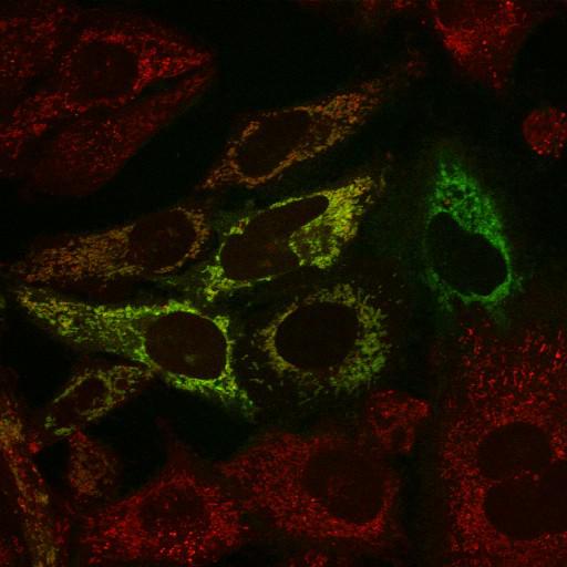
PINK1-YFP R98F (green) is found localized to mitochondria (red) in the presence of the mitochondrial depolarizing agent CCCP (carbonyl cyanide-m-chlorophenyl hydrazone). The R98F mutation in PINK1-YFP partially inhibits proteolytic cleavage. HeLa cells expressing PINK1-YFP R98F were stained with 10nM MitoTracker Red for 30 min to see mitochondrial morphology and then treated with 10µM CCCP for 3 hours. Cells were imaged using an inverted microscope (LSM510 Meta; Carl Zeiss, Inc.) with a 63× 1.4 NA oil differential interference contrast Plan Apo objective. Image contrast and brightness were adjusted in the accompanying image browser (LSM; Carl Zeiss, Inc.). This image corresponds to Fig 3d, R98F/CCCP bottom panels in J Cell Biol. 191: 933-942, 2010. Images in Fig3d include CIL#s 13713, 13714, 13715, 13716.

Biological Sources
NCBI Organism Classification
Homo sapiens
Cell Type
epithelial cell
cervical carcinoma
Cell Line
HeLa
Cellular Component
mitochondrion
mitochondrial inner membrane
mitochondrial outer membrane
Attribution
Names
Seok Min Jin
Michael Lazarou
Chunxin Wang
Lesley A. Kane
Derek P. Narendra
Richard J. Youle
Published
J Cell Biol. 191: 933-942, 2010.
Pubmed
21115803
Link
http://jcb-dataviewer.rupress.org/jcb/browse/...
Citation
Digital Object Identifier (DOI)
doi:10.7295/W9CIL13714
Archival Resource Key (ARK)
ark:/b7295/w9cil13714
Grouping This image is part of a group.
Imaging
Image Type
recorded image
Image Mode
single-spot confocal microscopy
Parameters Imaged
fluorescence emission
Source of Contrast
distribution of a specific protein
compartmentalization of stain or label
Visualization Methods
EYFP
probe for mitochondria
CMX rosamine (Mitotracker Red)
Processing History
brightness and contrast adjusted
Data Qualifiers
raw, unprocessed data
suitable for spatial measurements
Sample Preparation
Methods
living tissue
Relation To Intact Cell
dispersed cells in vitro
Dimensions
Spatial Axis Image Size Pixel Size
X 512px 0.279µm
Y 512px 0.279µm