Alternate header for print version


Licensing
Attribution Non-Commercial Share Alike:This image is licensed under a Creative Commons Attribution, Non-Commercial Share Alike License. View License Deed | View Legal Code
tweet  
*CIL – Cell Image Library accession number. Please use this to reference an image.

CIL:13724*  Cite 
Description

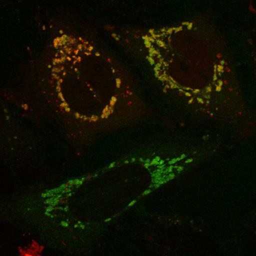
Expression of R98F PINK1-YFP (green) induces mitochondrial translocation of mCherry-Parkin (red) in the presence of the mitochondrial depolarizing agent CCCP (carbonyl cyanide-m-chlorophenyl hydrazone), indicating that this PINK1 mutant is functional for inducing Parkin translocation when located on the outer michondrial membrane. The R98F mutation in PINK1-YFP partially inhibits proteolytic cleavage. Transfected HeLa cells were treated with CCCP (10 µM) for 1 hr. Live cell imaging was performed on an LSM510 Meta (Carl Zeiss, Inc) with a 63x 1.4 NA oil differential interference contrast Plan Apo objective. Image contrast and brightness were adjusted in the LSM image browser (Zeiss). This image corresponds to Fig 4e, bottom row, with DMSO control in Fig 4e, top row of J Cell Biol, 191: 933-942, 2010. Images in Fig 4 include CIL#s 13733, 13734, 13729, 13730, 13731, 13732, 13717, 13718, 13719, 13720, 13721, 13722, 13723, 13724.

Biological Sources
NCBI Organism Classification
Homo sapiens
Cell Type
endothelial cell
Cell Line
HeLa
cervical carcinoma
Cellular Component
mitochondrion
cytosol
nucleus
mitochondrial inner membrane
mitochondrial outer membrane
Attribution
Names
Seok Min Jin
Michael Lazarou
Chunxin Wang
Lesley A. Kane
Derek P. Narendra
Richard J. Youle
Published
J Cell Biol, 191: 933-942, 2010.
Pubmed
21115803
Link
http://jcb-dataviewer.rupress.org/jcb/browse/...
Citation
Digital Object Identifier (DOI)
doi:10.7295/W9CIL13724
Archival Resource Key (ARK)
ark:/b7295/w9cil13724
Grouping This image is part of a group.
Imaging
Image Type
recorded image
Image Mode
single-spot confocal microscopy
Parameters Imaged
fluorescence emission
Source of Contrast
distribution of a specific protein
Visualization Methods
EYFP
mCherryFP
Processing History
brightness and contrast adjusted
Data Qualifiers
raw, unprocessed data
suitable for spatial measurements
Sample Preparation
Methods
living tissue
Relation To Intact Cell
dispersed cells in vitro
Dimensions
Spatial Axis Image Size Pixel Size
X 512px 0.186µm
Y 512px 0.186µm