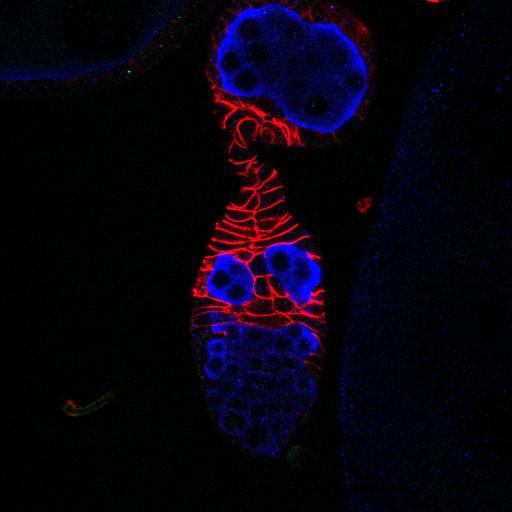
boi controls follicle stem cell proliferation. Germaria from boi[e] mutant in which germ cells (blue, anti-Vasa) and differentiating follicle cells (red, anti-Fas3) are labeled. Stalks between the germarium and the first budded egg chamber average 9 cells in wild type (CIL#13735) and 18 cells in boi[e] mutants (see also CIL# 13737). boi mutants exhibit defects in cyst packaging (indicated by side-by-side cysts in a single plane or two cysts surrounded by a single epithelium, and polarization defects (round cells, changes in Fas3 staining). Image correlates to Fig 1D in J Cell Biol. 2010. 191: 943-952.
Fly ovaries were dissected and fixed as described previously (O’Reilly et al., 2008). Primary antibodies were 1:2,000 rabbit anti-Vasa (Hay et al., 1990); 1:25 mouse anti-Fas3 (Developmental Studies Hybridoma Bank [DSHB]. Secondary antibodies used were either FITC, Cy3, and/or Cy5 conjugated to species-specific secondary antibodies (Jackson ImmunoResearch Laboratories, Inc.). Samples were mounted in Vectashield mounting medium. Images were collected at room temperature (22C) using 40× (1.25 NA) or 63× (1.4 NA) oil immersion lenses (Leica) on an upright microscope (DM 5000; Leica) coupled to a confocal laser scanner (TCS SP5; Leica). LAS AF SP5 software (Leica) was used for data acquisition. Images representing individual channels of single confocal slices from the center of each germarium were exported as TIFF files, and images were converted to figures using Photoshop software (Adobe).
| Spatial Axis | Image Size | Pixel Size |
|---|---|---|
| X | 512px | 0.2921µm |
| Y | 512px | 0.2921µm |
| Z | 1px | 0.042µm |