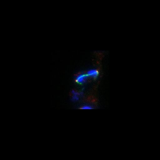
3HA-Bfa1 (red) localizes to both spindle pole bodies (SPBs) in anaphase in dyn1Δ cells with constitutive targeting of Tem1 to both SPBs. However, the constitutive presence of Tem1 on the SPBs impaired the spindle position checkpoint in the dyn1Δ cells. Anaphase was determined by spindle morphology (tubulin, green) and nuclear morphology (DAPI, blue). A DIC image is also shown (hidden). Image is Fig S3 in J Cell Biol. (2011) 192: 599-614.
Cells (MATa tem1::KanMX ura3::eGFP-CNM67–TEM1::URA3 dyn1::URA3 bfa1::3HA-BFA1) grown in rich media at 14C for 24 hours were fixed for 15 min in 3.7% formaldehyde and 0.1 M potassium phosphate buffer, pH 6.4. Cells were then washed twice with 0.1 M potassium phosphate buffer, pH 6.4, and resuspended in 1.2 M sorbitol in 0.12 M K2HPO4/0.033 M citric acid, pH 5.9. Fixed cells were digested with 0.1 mg/ml zymolyase-100T (US Biological) and 1/10 volume of glusulase (PerkinElmer) at 30C for 15 min, washed once, and resuspended in 1.2 M sorbitol in 0.12 M K2HPO4/0.033 M citric acid, pH 5.9. Primary antibodies were anti-HA monoclonal antibody (HA.11; 1:500) and anti-tubulin (Abcam; 1:500). Secondary antibodies were: anti-mouse Cy3 (for HA) and anti-rat FITC (for tubulin). Cells were resuspended in DAPI (1 mg/ml). Imaging was performed at 25C using a Leica DM6000 microscope equipped with a 100x/1.40 NA oil immersion objective lens, A4, L5, and TX2 filters, and a digital CCD camera (DFC350, Leica). Pictures were processed with LAS AF (leica) and ImageJ software.
| Spatial Axis | Image Size | Pixel Size |
|---|---|---|
| X | 187px | 0.0642µm |
| Y | 187px | 0.0642µm |