Alternate header for print version


Licensing
Public Domain: This image is in the public domain and thus free of any copyright restrictions. However, as is the norm in scientific publishing and as a matter of courtesy, any user should credit the content provider for any public or private use of this image whenever possible. Learn more
tweet  
*CIL – Cell Image Library accession number. Please use this to reference an image.

CIL:14483*  Cite 
Description

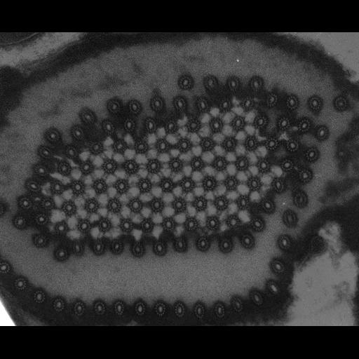
Electron micrograph of a thin section of fixed, embedded stained cells of Peranema trichophorum. Shown is a section of the "rodorgan', a bundle of 13-protofilment microtubules with pseudo-hexagonal symmetry. The section thickness was approximately 400 angstroms(silver grey section). Image recorded on Kodak SO-163 film, developed in straight D-19 developer, scanned on a Perkin-Elmer Model 1010G microdensitometer with a step size of 25 microns. Original electron-optical magnification was 46000. Sample was fixed in 2.5% glutaraldehyde, stained with Osmium tetroxide, dehydrated, embedded in Spurr's epoxy resin, sectioned, then stained with uranyl acetate and lead citrate. The rodorgan is thought to be used by Peranema for disrupting the microorganisms that are its prey. Unpublished micrograph

Biological Sources
NCBI Organism Classification
Peranema trichophorum
Cell Type
protozoa
Cellular Component
microtubule
interphase microtubule organizing center
Attribution
Names
John Murray
Citation
Digital Object Identifier (DOI)
doi:10.7295/W9CIL14483
Archival Resource Key (ARK)
ark:/b7295/w9cil14483
Imaging
Image Type
transmission electron microscopy (TEM)
Parameters Imaged
electron density
Source of Contrast
differences in adsorption or binding of stain
Visualization Methods
uranyl salt
lead salt
osmium tetroxide
Data Qualifiers
raw, unprocessed data
suitable for spatial measurements
Dimensions
Spatial Axis Image Size Pixel Size
X 1440px 0.49nm
Y 1180px 0.49nm