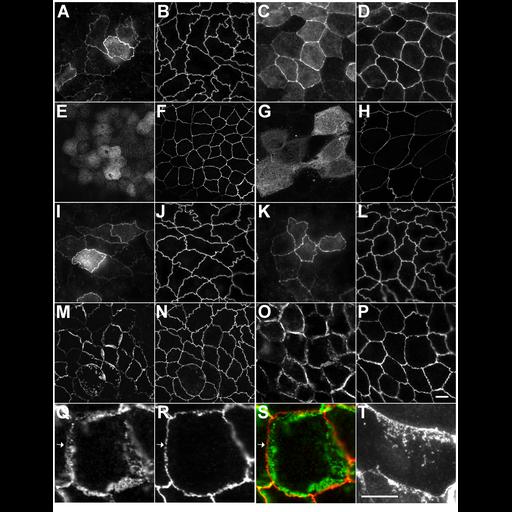
The unique-5 (U5) and unique-6 (U6) motifs of the MAGUK protein, ZO-1 are required for localization of ZO-1 to the apical tight junction. Cell lines expressing the myc-tagged transgenes ZO1-FL (A and B), ZNU6 (aa1-888) (C and D), ZNS (aa1-584) (E and F), deltaU5 (G and H), deltaSH3 (I and J), deltaGUK (K and L), deltaU6 (M and N), and ZN (aa1-806) (O and P) were stained with an antisera against c-myc (A, C, E, G, I, K, M, O, Q, and T) and antibodies specific for the endogenous ZO-1 (B, D, F, H, J, L, N, P, and R). The distribution of constructs lacking the U6 motif was strikingly different from those lacking the other conserved domains. The center field in O and P is magnified 2.6-fold in Q and R, respectively, and are merged in S (green, myc; red, endogenous ZO-1). At high magnification, ZO-1 lacking U6 staining is in linear strands that looped and wandered several micrometers from the apical junctinoal complex. (T) Another high magnification image of the ectopic strands observed in U6 cells. All images obtained by wide-field microscopy except E, F, G, H, M, and N, which were reconstructed from 5.0–8.0-µm-thick confocal stacks. Bar, 10.0 µm in A–P, and 4.0 µm in Q–T.
MDCK-II tet off cells were plated on glass coverslips at a low density (1.0 x 10[4] cells/ml) and incubated in the absence of doxycyclin for 4 d before processing. Cells were fixed in 1% paraformaldehyde, permeabilized in 0.2% Triton X-100 and incubated in anti-myc antibody and rat anti-ZO1 hybridoma supernatant (R40.76) followed by Cy3-conjugated donkey anti-rat secondary antibody and a secondary antibody specific for anti-myc. Cells were mounted in Mowiol with 1.0% n-propyl gallate. Wide-field images were acquired on a Nikon E800 microscope using 60x or 100x Plan Apo lenses and an Orca ER cooled CCD camera controlled with the Metamorph Imaging software package. The special green filter set #C4607 was used for Cy3 images. Confocal images were acquired on a Zeiss LSM510 Meta using a 100x Plan Apo lens (Thornwood, NY). Confocal Stacks (E,F,G,H,M,N) and image projections were generated with Zeiss LSM Image Browser version 3.2. Contrast adjustment and montages were generated using Adobe Photoshop (version 6.0; San Jose, CA). Figure 6 in Mol Biol Cell (2006). 18:721-731.