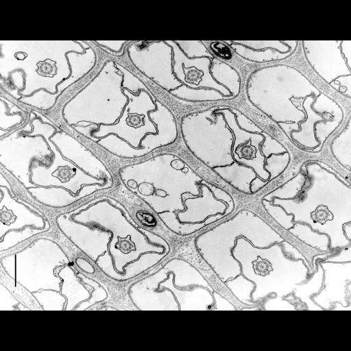
Paramecium multimicronucleatum is covered with cilia which cilia arise from regular surface depressions roughly rectangular in shape. This image is adapted with permission from Fig. 1 in J. Cell Biol. 49:1-20, 1971. The plasma membrane covers the exterior of the cell and this is closely attached to a mosaic of alveolar sacs internally which in this micrograph are expanded. Borders of adjacent alveolar sacs form septa along each row of cilia and sometimes laterally as well. Septa do not follow the crests of the ridges. The ridges of the rectangular depressions at their tips are filled internally with a granulo-fibrillar material. Trichocyst tips penetrate into the transverse margins along each row of cilia. In this micrograph the trichocysts have been expelled and in a few cases only their membranous casings remain. Collidine buffered fixative. TEM taken on 12/20/68 by R. Allen with Philips 300 operating at 60kV. Neg. 16,000X. Bar = 0.5µm. The negative was printed to paper and the image was scanned to Photoshop. This digitized image is available for qualitative analysis. An unprocessed, high resolution version of this image (CIL:12029) is in the library and available for quantitative analysis. Standard glutaraldehyde fixation followed by osmium tetroxide, dehydrated in alcohol and embedded in an epoxy resin. Microtome sections prepared at approximately 75nm thickness. Additional information available at (http://www5.pbrc.hawaii.edu/allen/).
| Spatial Axis | Image Size | Pixel Size |
|---|---|---|
| X | 2793px | —— |
| Y | 2154px | —— |