Alternate header for print version


Licensing
Public Domain: This image is in the public domain and thus free of any copyright restrictions. However, as is the norm in scientific publishing and as a matter of courtesy, any user should credit the content provider for any public or private use of this image whenever possible. Learn more
tweet  
*CIL – Cell Image Library accession number. Please use this to reference an image.

CIL:191*  Cite 
Description

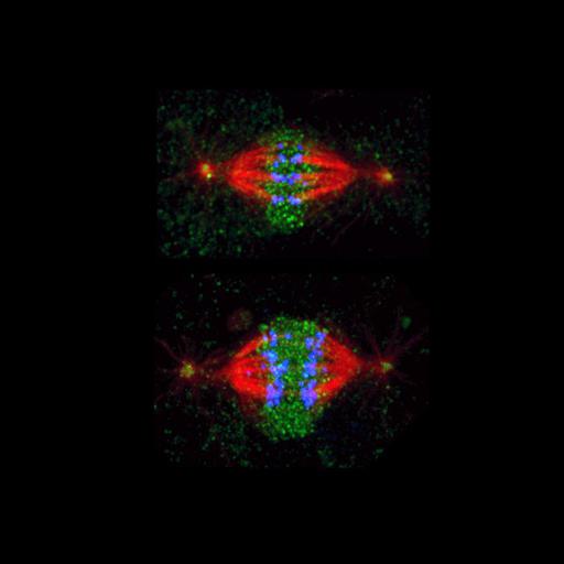
The image shows mitotic metaphase (upper) and anaphase (lower) in Drosophila tissue culture cells immunostained for the microtubule severing protein katanin (green), microtubules (red) and kinetochores/chromosomes (blue). Katanin targets to chromosomes during both metaphase and anaphase and is responsible for inducing the depolymerization of attached microtubule plus-ends.

Technical Details

See D. Zhang et al. 2007 Three microtubule severing enzymes contribute to the "Pacman-flux" machinery that moves chromosomes. J Cell Biol 177, 231-242.

Biological Sources
NCBI Organism Classification
Drosophila melanogaster
Cell Line
S2
Cellular Component
spindle
nuclear chromosome
Biological Context
Biological Process
mitosis
Attribution
Names
David Sharp
Dong Zhang
Gregory Rogers
Daniel Buster
Citation
Digital Object Identifier (DOI)
doi:10.7295/W9CIL191
Archival Resource Key (ARK)
ark:/b7295/w9cil191
Imaging
Image Type
recorded image
Image Mode
spinning disk confocal microscopy
Parameters Imaged
fluorescence emission
Source of Contrast
distribution of epitope
Processing History
montage
Data Qualifiers
processed data
Sample Preparation
Methods
methanol fixed tissue
Relation To Intact Cell
whole mounted tissue
Dimensions
Spatial Axis Image Size Pixel Size
X 700px ——
Y 523px ——