Alternate header for print version


Licensing
Attribution Non-Commercial Share Alike:This image is licensed under a Creative Commons Attribution, Non-Commercial Share Alike License. View License Deed | View Legal Code
tweet  
*CIL – Cell Image Library accession number. Please use this to reference an image.

CIL:21573*  Cite 
Description

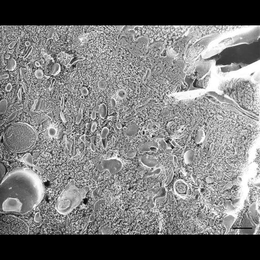
Vesicles and early endosomes under the membranelles of the peniculus of the buccal cavity. Cilia and basal bodies are tangentially fractured in this quick-freeze deep-etch preparation. Preendosomal vesicles, clathrin-coated and partially uncoated, that arise from the clathrin-coated ends of parasomal sacs lie nearest the basal body/cilium complexes. Internal to these are the many early endosomes to which the uncoated preendosomal vesicles fuse and from which smaller clathrin-coated vesicles bud off. Blebs protrude from one early endosome. Carrier vesicles of about 100nm in diameter are found internal to the early endosomes. TEM taken on 7/19/88 by C. Schroeder with Zeiss 10A operating at 80kV. Neg. 12,000X. Bar = 0.5µm. Adapted with permission from J. Cell Sci. A print of the negative was scanned and processed in Photoshop. This image is best used for qualitative analysis. A high resolution image (CIL:12636) is available for quantitative analysis. Additional information available at (http://www5.pbrc.hawaii.edu/allen/).

Biological Sources
NCBI Organism Classification
Paramecium multimicronucleatum
Cell Type
cell by organism
eukaryotic cell
Eukaryotic Protist
Ciliated Protist
Cellular Component
cytoplasm
early endosome
clathrin coat of coated pit
vesicle membrane
Attribution
Names
Richard Allen (University of Hawaii)
Citation
Digital Object Identifier (DOI)
doi:10.7295/W9CIL21573
Archival Resource Key (ARK)
ark:/b7295/w9cil21573
Grouping This image is part of a group.
Imaging
Image Type
transmission electron microscopy (TEM)
illumination by electrons
Image Mode
electron density
Parameters Imaged
elastic scattering of electrons
electron density
elevation
Source of Contrast
differences in deposition of metal shadow
Visualization Methods
transmission electron microscopy (TEM)
Processing History
freeze_fracture/freeze_etch
shadowing and plating
recorded image
film
Print of negative scanned for Photoshop
Data Qualifiers
processed data
Dimensions
Spatial Axis Image Size Pixel Size
X 2658px ——
Y 2141px ——