Alternate header for print version


Licensing
Public Domain: This image is in the public domain and thus free of any copyright restrictions. However, as is the norm in scientific publishing and as a matter of courtesy, any user should credit the content provider for any public or private use of this image whenever possible. Learn more
tweet  
*CIL – Cell Image Library accession number. Please use this to reference an image.

CIL:21642*  Cite 
Description

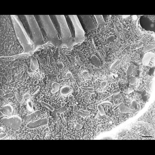
The largest accumulation of endosomal vesicles and early endosomes is next to the buccal cavity area under the membranelles of the peniculi and quadrulus. Cilia of the peniculi have a very close packing but have only three rows of parasomal sacs, two rows on the two margins of the light rows of cilia and a third row down the middle of the eight ciliary rows separating the dorsal from the ventral peniculi. Early endosomes have smooth membranes except for their clathrin coated evaginations waiting to be pinched off. In this micrograph most membranes are uncoated. Two tangentially fractured basal bodies showing the globular masses seen in thin sections in their shafts are embedded in strands of the meshwork of the filamentous reticulum. Only one cilium shows the P-fracture face of the ciliary membrane, all others appear not to have been fractured. Rings of IMPs or transmembrane particles are seen at ciliary bases. TEM taken on 6/4/91 by R. Allen with Zeiss 10A operating at 80kV. Neg. 19,800X. Bar = 0.25µm. A print of the negative was scanned and processed in Photoshop. This image is best used for qualitative analysis. A high resolution image (CIL:12635) is available for quantitative analysis. Additional information available at (http://www5.pbrc.hawaii.edu/allen/).

Biological Sources
NCBI Organism Classification
Paramecium multimicronucleatum
Cell Type
cell by organism
eukaryotic cell
Eukaryotic Protist
Ciliated Protist
Cellular Component
endosome
cilium
oral apparatus
Biological Context
Biological Process
endosome organization
oral apparatus organization
Attribution
Names
Richard Allen (University of Hawaii)
Citation
Digital Object Identifier (DOI)
doi:10.7295/W9CIL21642
Archival Resource Key (ARK)
ark:/b7295/w9cil21642
Grouping This image is part of a group.
Imaging
Image Type
transmission electron microscopy (TEM)
illumination by electrons
Image Mode
electron density
Parameters Imaged
elastic scattering of electrons
electron density
elevation
Source of Contrast
differences in deposition of metal shadow
Visualization Methods
transmission electron microscopy (TEM)
Processing History
freeze_fracture/freeze_etch
shadowing and plating
recorded image
film
Print of negative scanned for Photoshop
Data Qualifiers
processed data
Dimensions
Spatial Axis Image Size Pixel Size
X 2670px ——
Y 2154px ——