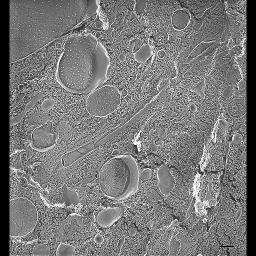
Quick-freeze deep-etch view of a radial arm showing a collecting canal surrounded with smooth spongiome as well as a few decorated tubules. However several larger vesicles lie next to the radial arm containing a lumen resembling the contractile vacuole. These vesicles appear to be rounded decorated tubules which have been transformed into vesicles or have not yet tubulated and become part of the decorated spongiome of the mature radial canal. Although we cannot be certain without further study we believe this may represent a dividing cell as we do know that the radial arms of the dividing cell, both their smooth and decorated spongiomes, shorten as the new CVCs develop (Allen et al., J. Cell Sci. 96:469-475, 1990; Fok et al., J. Eukaryot. Microbiol. 49:185-196, 2002. The P-face of these vesicles has a large number of IMPs like that seen in collecting canals. The highly porous E-face of several vesicles indicates that these vesicles are indeed products of the decorated spongiome. TEM taken on 6/22/88 by C. Schroeder with Zeiss 10A operating at 80kV. Neg. 9,780X. Bar = 0.5µm. A print of the negative was scanned and processed in Photoshop. This image is best used for qualitative analysis. A high resolution image (CIL:13130) is available for quantitative analysis. Additional information available at (http://www5.pbrc.hawaii.edu/allen/).
| Spatial Axis | Image Size | Pixel Size |
|---|---|---|
| X | 1758px | —— |
| Y | 1902px | —— |