Alternate header for print version


Licensing
Public Domain: This image is in the public domain and thus free of any copyright restrictions. However, as is the norm in scientific publishing and as a matter of courtesy, any user should credit the content provider for any public or private use of this image whenever possible. Learn more
tweet  
*CIL – Cell Image Library accession number. Please use this to reference an image.

CIL:252*  Cite 
Description

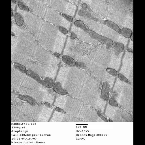
Thin section electron microscopy of diaphragm (skeletal) muscle from a wild type mouse. Sample was viewed with a Hitachi 7600 electron microscope (accelerating voltage 80 KV) and imaged with an AMT digital camera. Image taken at 30,000X magnification. This image is part of a set of wild type diaphragm muscle images that served as controls for the study of the delta-sarcoglycan knockout mouse. Both the set of wild-type and delta-sarcoglycan knockout mouse images are part of The CELL: an image library. Reference: Nat Med. 2008 Apr;14(4):442-7. Genetic and pharmacologic inhibition of mitochondrial-dependent necrosis attenuates muscular dystrophy. Millay DP, Sargent MA, Osinska H, Baines CP, Barton ER, Vuagniaux G, Sweeney HL, Robbins J, Molkentin JD (tissue preparation described in Fewell JG, et al. A treadmill exercise regimen for identifying cardiovascular phenotypes in transgenic mice. Am J Physiol. 1997;273:H1595–H1605)

Biological Sources
NCBI Organism Classification
Mus musculus
Cell Type
skeletal muscle cell
Cellular Component
dystroglycan complex
Biological Context
Biological Process
plasma membrane organization
Molecular Function
structural molecule activity
Attribution
Names
Hanna Osinska
Pubmed
18345011
Citation
Digital Object Identifier (DOI)
doi:10.7295/W9CIL252
Archival Resource Key (ARK)
ark:/b7295/w9cil252
Grouping This image is part of a group.
Imaging
Image Type
recorded image
Image Mode
transmission electron microscopy (TEM)
Parameters Imaged
electron density
Source of Contrast
uranyl salt
lead salt
Visualization Methods
detection of electrons
Processing History
unprocessed raw data
Sample Preparation
Methods
glutaraldehyde fixed tissue
osmium tetroxide fixed tissue
Relation To Intact Cell
microtome-sectioned tissue
Dimensions
Spatial Axis Image Size Pixel Size
X 1696px 3.03nm
Y 2016px 3.03nm