Alternate header for print version


Licensing
Public Domain: This image is in the public domain and thus free of any copyright restrictions. However, as is the norm in scientific publishing and as a matter of courtesy, any user should credit the content provider for any public or private use of this image whenever possible. Learn more
tweet  
*CIL – Cell Image Library accession number. Please use this to reference an image.

CIL:25314*  Cite 
Description

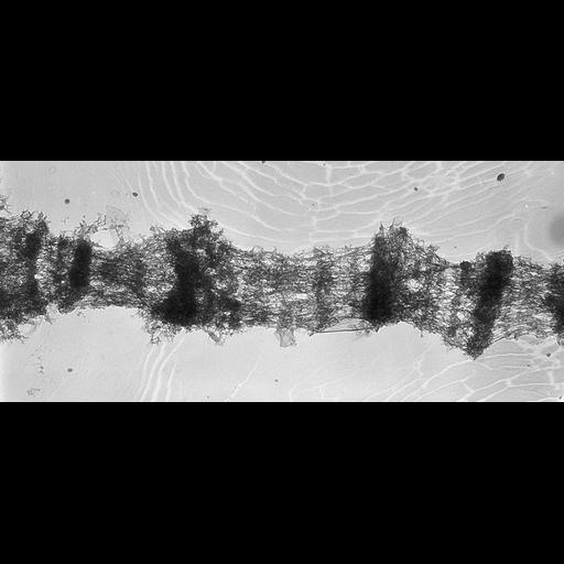
Fat body cells from larvae of Drosophila melanogaster were dissected, fixed with formalin-acetic acid, and squashed on a glass slide. After freezing in liquid nitrogen, the cover slips removed, and the slide thawed and stained with 1% uranyl acetate, rinsed in 50% ethanol, dehydrated, transferred to amyl acetate, and covered with 2% parlodion in amyl acetate. Selected areas of the parlodion film, are transferred to formvar coated EM grids, and the parlodion removed by immersion in amyl acetate (see Ris H, 1981 Stereoscopic Electron Microscopy of Chromosomes, Meth Cell Biol 22:77-96). Grids were observed using the Madison High Voltage Electron Microscope at 1 MeV. Image shows a series of bands and interbands in a polytene chromosome. This image was taken with a tilt angle of 55 degrees. The accompanying grouped image, taken at 45 degrees forms a stereo pair, providing an oblique 3D view of the chromosome.

Biological Sources
NCBI Organism Classification
Drosophila melanogaster
Cell Type
fat body cell
Cellular Component
polytene chromosome
Attribution
Names
Hans Ris
Citation
Digital Object Identifier (DOI)
doi:10.7295/W9CIL25314
Archival Resource Key (ARK)
ark:/b7295/w9cil25314
Grouping This image is part of a group.
Imaging
Image Type
film
Image Mode
transmission electron microscopy (TEM)
Parameters Imaged
elastic scattering of electrons
Source of Contrast
differences in adsorption or binding of stain
Visualization Methods
uranyl salt
Processing History
unprocessed raw data
Sample Preparation
Methods
formaldehyde fixed tissue
acetic acid treatment
Relation To Intact Cell
squashed preparation
Dimensions
Spatial Axis Image Size Pixel Size
X 4030px 1nm
X 1727px 1nm