Alternate header for print version


Licensing
Attribution Non-Commercial Share Alike:This image is licensed under a Creative Commons Attribution, Non-Commercial Share Alike License. View License Deed | View Legal Code
tweet  
*CIL – Cell Image Library accession number. Please use this to reference an image.

CIL:26573*  Cite 
Description

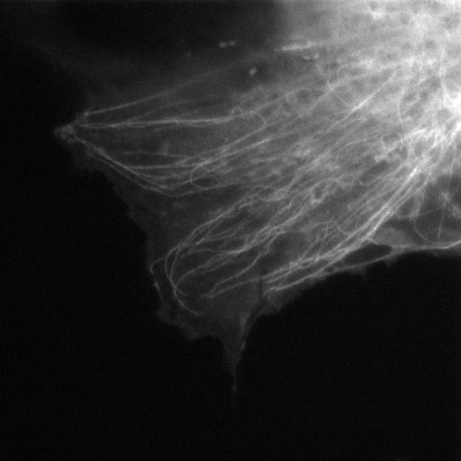
Movie raw file of RPE-1 cells expressing GFP-alpha-tubulin and depleted for pVHL (Von Hippel-Lindau) by ectopic expression of shRNAmir against VHL. Video used to manually track MTs and determine growth speed and shrinkage time. Cells on round coverslips were transferred to a homemade holding device, and 600 µl of the appropriate medium was added. The video was acquired with a microscope (IX70 Delta Vision Spectris; Olympus), temperature-controlled at 37C, using a 100× NA 1.4 differential interference contrast (DIC) oil Plan-Apochromat objective, ex 470 em 520, and a camera (CoolSNAP HQ; Roper Industries) with an exposure time of 200 ms and a frame rate of 2Hz. Acquisition software used was SoftWoRx version 3.3.4 (Applied Precision). This video corresponds to J Cell Biol. 2010. 190: 991-1003--Supplementary Figure 1.

Biological Sources
NCBI Organism Classification
Homo sapiens
Cell Type
renal pigment epithelium
epithelial cell
Cell Line
RPE-1
Cellular Component
microtubule
cytoskeleton
protein complex
Biological Context
Biological Process
protein polymerization
Molecular Function
GTPase activity
structural molecule activity
Attribution
Names
Claudio R. Thoma
Alexandre Matov
Katrin L. Gutbrodt
Christian R. Hoerner
Zlatko Smole
Wilhelm Krek
Gaudenz Danuser
Published
J Cell Biol. 2010. 190: 991-1003.
Pubmed
20855504
Link
http://jcb-dataviewer.rupress.org/jcb/browse/...
Citation
Digital Object Identifier (DOI)
doi:10.7295/W9CIL26573
Archival Resource Key (ARK)
ark:/b7295/w9cil26573
Grouping This image is part of a group.
Imaging
Image Type
recorded image
Image Mode
widefield illumination
time lapse microscopy
Parameters Imaged
fluorescence emission
Source of Contrast
distribution of a specific protein
Visualization Methods
EGFP
Processing History
unprocessed raw data
Data Qualifiers
raw, unprocessed data
suitable for spatial measurements
Sample Preparation
Methods
living tissue
Relation To Intact Cell
dispersed cells in vitro
Dimensions
Spatial Axis Image Size Pixel Size
X 1024px 0.0663µm
Y 1024px 0.0663µm
Time 0.5 seconds 151