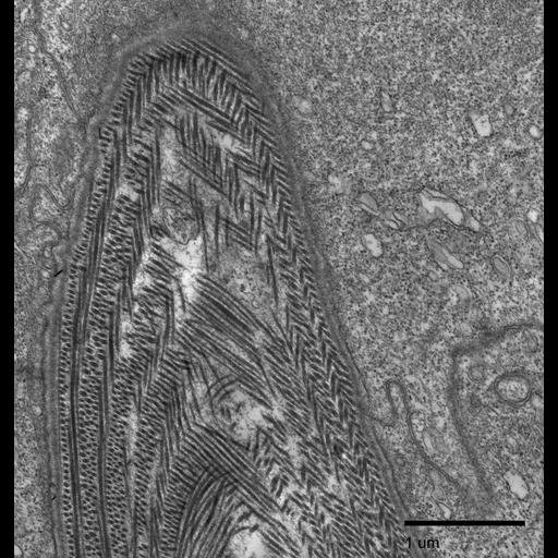
Collagen fibrils in the basal plate of Fundulus heteroclitus scales. The basal plate is made of thick collagen fibrils about 100 nm in diameter, which are organized into a plywood-like structure. Note the changing orientation of the collagen fibrils at every layer of fibrils. During formation of this plywood-like organization, collagen fibrils are synthesized in a direction that is approximately perpendicular to the preceding ply. These well-defined layers of collagen constitute the stratum compactum of the dermis of Fundulus heteroclitus.
Fundulus heteroclitus scales were chemically fixed with 2.5% glutaraldehyde, 2% formaldehyde in 0.1M cacodylate buffer (pH 7.3), then post-fixed in 4% osmium tetroxide and stained en bloc in 1% uranyl acetate. The scales were then dehydrated in a graded series of ethanol and infiltrated with Spurr’s resin. Thin sections of 70 nm were trimmed using a diamond knife and post-stained in uranyl acetate and lead citrate. This micrograph was imaged using a Phillips CM 100 transmission electron microscope at an accelerating voltage of 80 kV.
| Spatial Axis | Image Size | Pixel Size |
|---|---|---|
| X | 1689px | 2.59nm |
| Y | 1781px | 2.59nm |