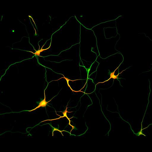
Development of the axon and dendritic arbors in cultured hippocampal neurons after 5 days in vitro. MAP2 staining (red) highlights the dendrites, while microtubule staining (green) reveals both the axons and dendrites in the field of view. Neurons at 2, 3, 5 and 7 days in vitro are represented in this image group. Detailed methods: Embryonic rat hippocampal neurons were prepared as previously described (see Kaech and Banker, 2006, Nat Protoc). Cells were prepared for fluorescent staining as previously described (Withers and Banker, 1998, in Culturing Nerve Cells, MIT Press). Briefly, cells were fixed (4% formaldehyde, 4% sucrose in phosphate buffered saline, pH 7.4), permeabilized with 0.25% Triton and immunostained for tubulin (monoclonal DM1A, from Sigma with Alexa 488 conjugated secondary, excitation, 494, emission, 519 [Invitrogen, Molecular Probes]) and MAP2 (polyclonal Ab266, from S. Halpain with d549 conjugated secondary, excitation, 555, emission, 568, Jackson Immunoresearch). Images were acquired with a Leica DMRA microscope with a 20X (Fluotar, NA 0.5) lens, Photometrics CoolSnap ES CCD camera and MetaMorph software. Image generated with the MetaMorph color combine function.
| Spatial Axis | Image Size | Pixel Size |
|---|---|---|
| X | 1300px | 0.339µm |
| Y | 1030px | 0.339µm |