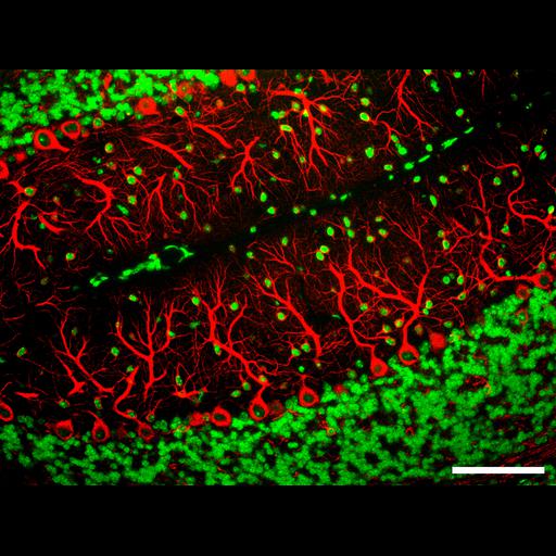
Immunohistochemistry of neuron-specific β-III-tubulin (red) shows the delicate dendrites of Purkinje cells in the cerebellum. Cell nuclei are revealed with DAPI staining (blue). Scale bar, 100 µm.
The adult mouse were perfused transcardially with 4% PFA following anesthetization with avertin. The brain was dissected out and fixed by immersion in 4% paraformaldehyde (PFA) overnight at 4 °C, embedded in paraffin and cut into 5-µm-thick sagittal sections, which were deparaffinized using a standard histology protocol immediately before immunohistochemical staining. In the staining procedure, tissue sections were permeabilized with 0.5% Triton X-100 (Sigma) for 10 minutes, then blocked with the buffer containing 10% normal goat serum (Sigma), 1% (w/v) bovine serum albumin (BSA) and 0.2% (v/v) Triton X-100 for 2 hours at RT, followed by incubation with anti-βIII-tubulin monoclonal antibody (Covance) at1:50 dilution in the dilution buffer (2% normal goat serum, 1% BSA, 0.1% Triton X-100) overnight at 4 °C. Samples were subsequently incubated with FITC- and/or Cy3-conjugated species-specific secondary antibody/antibodies in the dilution buffer (1:200 dilution) for 1 hour at RT. VECTASHIELD Mounting Medium with DAPI (Vector Laboratories) was used to mount the fluorescently labeled samples and to stain cell nuclei. Images were digitally acquired using a fluorescence microscope (Nikon Eclipse 660) equipped with Spot cooled CCD camera (Diagnostic Instruments).
| Spatial Axis | Image Size | Pixel Size |
|---|---|---|
| X | 4667px | 7.77µm |
| Y | 3500px | 7.77µm |