Alternate header for print version


Licensing
This image is copyright protected. Any public or private use of this image is subject to prevailing copyright laws. Please contact the content provider of this image for permission requests.
tweet  
*CIL – Cell Image Library accession number. Please use this to reference an image.

CIL:35124*  Cite 
Description

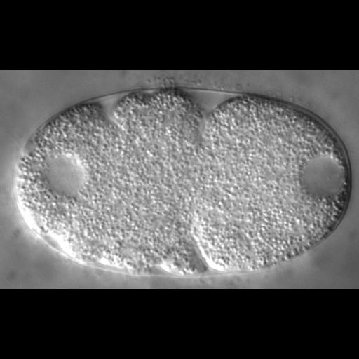
This time lapse series illustrates the early division up to the four-cell stage of a wild-type C. elegans embryo by DIC microscopy. The first C. elegans embryonic division of the P0 zygote is asymmetric and generates an anterior AB cell, and a smaller posterior P1 cell. These cells have different developmental fates and division timing, with AB dividing approximately 2 min before P1. Additionally, in early C. elegans embryonic cell cycles, S and M phases rapidly alternate without apparent gap phases. The series corresponds to experiments shown in Fig. 1B and Movie 2 of Mouysset et al. Movie is CIL 25629 and original data file is CIL 35124.

Technical Details

Eggs were extruded in M9 buffer from dissected adult worms and mounted on 2% agarose pads. Recordings were acquired at 4-s intervals with AxioCam HR or AxioCam MRc cameras mounted on Axioplan2 Imaging or Axiophot microscopes, respectively, equipped with differential interference contrast (DIC) optics (100×/1.3 Plan-Neofluar; Carl Zeiss).

Biological Sources
NCBI Organism Classification
Caenorhabditis elegans
Cell Type
early embryonic cell
Cell Line
N2
Cellular Component
nucleus
Biological Context
Biological Process
mitosis
cell division
nucleus organization
Molecular Function
ATPase activity
Attribution
Names
Julien Mouysset
Alexandra Deichsel
Sandra Moser
Carsten Hoege
Anthony A. Hyman
Anton Gartner
Thorsten Hoppe
Published
Proc Natl Acad Sci. 2008. 105: 12879-12884.
Pubmed
18728180
Link
Proceedings National Academy of Sciences USA
Citation
Digital Object Identifier (DOI)
doi:10.7295/W9CIL35124
Archival Resource Key (ARK)
ark:/b7295/w9cil35124
Grouping This image is part of a group.
Imaging
Image Type
recorded image
Image Mode
differential interference contrast microscopy
time lapse microscopy
Parameters Imaged
optical path length gradient
Source of Contrast
boundaries between regions with different refractive index
Processing History
unprocessed raw data
Data Qualifiers
raw, unprocessed data
Sample Preparation
Methods
living tissue
Relation To Intact Cell
whole mounted tissue
Dimensions
Spatial Axis Image Size Pixel Size
X 960px ——
Y 586px ——
Time 4 seconds 166