Alternate header for print version


Licensing
Public Domain: This image is in the public domain and thus free of any copyright restrictions. However, as is the norm in scientific publishing and as a matter of courtesy, any user should credit the content provider for any public or private use of this image whenever possible. Learn more
tweet  
*CIL – Cell Image Library accession number. Please use this to reference an image.

CIL:35127*  Cite 
Description

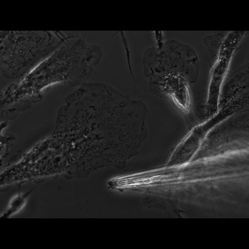
Primary bone marrow macrophages derived from mouse plated on a coverslip and imaged live. Phase contrast microscopy allows us to see the dense f-actin rich leading edge, ruffles, mitochondria, the nucleus, and other organelles throughout the volume of the cells. At the center of the image is a polarized lamellipod of a crawling cell. The glass micropipette at the bottom of the image is delivering CSF (colony stimulating factor) locally and the cell may be responding.

Technical Details

Live bone marrow macrophages plated in MatTek dish and imaged with a 60X N.A. 1.4 phase 3 Olympus objective on an IX71 microscope with a Cooke Sensicam QE camera and IPLab software running on a Dell Windows XP computer.

Biological Sources
NCBI Organism Classification
Mus musculus
Cell Type
macrophage
Cell Line
Bone marrow
Cellular Component
ruffle membrane
Biological Context
Biological Process
ruffle organization
Molecular Function
chemokine receptor activity
Mouse Gross Anatomy
TS24,femur
Attribution
Names
Michael Cammer
Dianne Cox
Citation
Digital Object Identifier (DOI)
doi:10.7295/W9CIL35127
Archival Resource Key (ARK)
ark:/b7295/w9cil35127
Grouping This image is part of a group.
Imaging
Image Type
recorded image
Image Mode
phase contrast microscopy
Parameters Imaged
elastic scattering of photons
Source of Contrast
differences in amount of elastic light scattering
Visualization Methods
visualization of contiguous regions
Processing History
unprocessed raw data
Data Qualifiers
raw, unprocessed data
suitable for spatial measurements
Sample Preparation
Methods
living tissue
Relation To Intact Cell
dispersed cells in vitro
Dimensions
Spatial Axis Image Size Pixel Size
X 1376px 0.11µm
Y 1040px 0.11µm