Alternate header for print version


Licensing
Public Domain: This image is in the public domain and thus free of any copyright restrictions. However, as is the norm in scientific publishing and as a matter of courtesy, any user should credit the content provider for any public or private use of this image whenever possible. Learn more
tweet  
*CIL – Cell Image Library accession number. Please use this to reference an image.

CIL:35167*  Cite 
Description

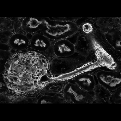
Dominant features of a nephron in the cortex of rat kidney showing f-actin localization in the glomerulus, a blood vessel, and proximal tubules. The glomerulus is the large ball at the left. The blood vessel that delivers blood to the glomerulus (Afferent arteriole) is the bright narrow tube shown both in elongated view and in cross section (upper right) that is lined with juxtaglomerular cells. The proximal convoluted tubules are cut to reveal the actin rich micro villi lining the interior surface. This thin optical section was acquired with a laser scanning confocal microscope and is a montage of a few adjacent fields imaged at high resolution.

Technical Details

Formaldehyde fixed rat kidney was cut by Vibratome and incubated with fluorescent labeled phalloidin, a toxin which binds to f-actin filaments. Imaging was performed using a BioRad MRC 600 laser scanning confocal microscope with a Kr/Ar laser. The microscope was a Nikon Diaphot with a 60X or 40X fixed tube length objective. Images of adjacent fields were manually stitched using Adobe Photoshop 2.5 on a Quadra Macintosh computer and may have manual contrast curves applied. Imaging performed at the Image Analysis Facility at the Albert Einstein College of Medicine.

Biological Sources
NCBI Organism Classification
Rattus rattus
Cell Type
podocyte (sensu Diptera)
epithelial cell
Cell Line
Cortex of kidney
Cellular Component
actin cytoskeleton
Biological Context
Biological Process
renal system process
Attribution
Names
Michael Cammer
Phyllis Novikoff
Citation
Digital Object Identifier (DOI)
doi:10.7295/W9CIL35167
Archival Resource Key (ARK)
ark:/b7295/w9cil35167
Imaging
Image Type
recorded image
Image Mode
single-spot confocal microscopy
fluorescence microscopy
Parameters Imaged
distribution of a specific protein
Source of Contrast
fluorescence emission
Visualization Methods
visualization of contiguous regions
fluorescent labels
Processing History
montage
Data Qualifiers
processed data
suitable for spatial measurements
Sample Preparation
Methods
chemically fixed tissue
vibratome-sectioned tissue
Relation To Intact Cell
chemically fixed tissue
Dimensions
Spatial Axis Image Size Pixel Size
X 1080px ——
Y 771px ——